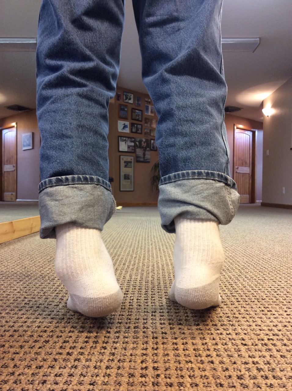
This Client went Phasic in their Gait. Do you know what that means ? We do, and so does McGill, Liebenson, Cook and many others.

Long ago on this blog we showed and discussed a video (link) that discussed Stu McGill's research of the human movements of Georges St-Pierre and David Loiseau. The basic tenets of that video were that the hips and shoulders are used for power production and that the spine-core are used for creating stiffness and stability for the ultimate power transmission through the limb.  He made it clear that if power is generated from the spine, it will suffer. 

Here on TGG we have long talked about phasic and antiphasic motions of the arms and shoulder-pelvic blocks during gait and locomotion/sport activity.  Many of our 1000+ blog writings and 80 podcasts have talked about spine pain and how spine pain clients reduce the antiphasic rotational (axial) nature of the shoulder girdle and pelvic girdle. In the video above, we see anything but antiphasic gait, to be clear, this is a classic representation of a phasic gait. This is pathologic gait, the frontal plane sway is exaggerated and necessary because there is no axial antiphasic motion.  There is essentially frozen arm and torso movements. This client has a long standing history of severe spine trauma and pain, their central pattern generators (CPG) had to make this motor pattern choice in an attempt to avoid pain and negotiate force streams across trauma zones. If you are curious and wish to go deeper down this rabbit hole, read the 30+ articles we have produced more specifically on arm swing and locomotor phasics, just click here.

In these types of cases, the client subconsciously makes the subcortial pattern choice (overrides the normal CPG) to rotate them as a solid unit to reduce spine rotation, axial loading and compression.  We could say that quite often spine pain disables the normal arm-leg pendulums via altering the shoulder-torso and hip-pelvis phasics and the CPG that dictates them. Normally, the spine and core must present sufficient amounts of recruited stiffness, yet mobility where necessary, to enable the locomotive power and velocity generated by movements of the shoulders and hips. These are the two main portals of limb movement off of the spine/core.  These principles holds true in gait and sport. For and interesting example, in human gait the psoas is not entirely a hip flexor initiator when it comes to leg swing, it is a huge hip flexion perpetuator. The initial hip flexion in human gait comes from derotating the obliqued pelvis, via abdominal contraction, on a stiff and stable spine.  Once the pelvis rotation is initiated, the femur can further pendulum forward (via contraction of the psoas and other muscles) on the forward accelerated pelvis in the hip joint proper creating an energy efficient movement (the towel flick/whip effect). This premise holds true in gait, running, kicking etc.  This is a solid principle of effective and efficient human locomotion. This principle also holds true for a punch or throwing an object, the stable torso/spine provides a stable anchor upon which to accelerate the arm in order to create a high velocity limb movement with power.  But here is where we get annoyed much of the time.  (Soap box Tangent coming up) How often do you read articles about tight ITBand, tight psoas, tight piriformis and the like ?  As a “diagnosis” these are weak and they are the “go to diagnosis or cause” of the unseasoned clinician, trainer, coach, therapist. If we all are to be really good at our job, we must go beyond what we see in someone’s gait (since it is the compensation) and go beyond the CNS neuroprotective strategy of tightness/shortness when there is weakness or motor pattern failure.  This does not mean that you cannot, or should not, incorporate restoration methods and principles to restore length-tension relationships in your client, it means you have to resolve ALL of the problems, including the aberrant CPG they have set up as a protective default to avoid injury or further injury. 

In the case above, returning the discussion to arm and leg swing, one must understand clearly that faulty arm swing patterns and lack of antiphasic torso and pelvis oscillation is a product of surgery,  trauma and more so, pain. The client is avoiding the antiphasic presentation (hence, he is phasic) for a reason and coaching more arm swing would be just about the dumbest intervention, so don’t be “that guy”. We know this is an altered motor pattern choice, not a new fixed set point. We know this because on clinical examination the range is available, we know because we examined for it, it is just not being used.  In an example of this same principle, in this case talking hip ranges of motion, McGill discusses the same in his paper*:

“Despite the large increases in passive hip ROM, there was no evidence of increased hip ROM used during functional movement testing. Similarly, the only significant change in lumbar motion was a reduction in lumbar rotation during the active hip extension maneuver (p < 0.05). These results indicate that changes in passive ROM or core endurance do not automatically transfer to changes in functional movement patterns. This implies that training and rehabilitation programs may benefit from an additional focus on grooving new motor patterns if newfound movement range is to be used.”

Think about that next time you stretch, or are stretched by someone. As we have said before, just because you increase someone’s range of motion, does not mean they will be able to incorporate that range of motion into a movement pattern, or compensation pattern for that matter. It is only ¼ of the equation: Range of Motion,  Skill (or proprioception),  Endurance (or the proportion of slow twitch muscle) and Strength (the proportion of fast twitch muscle). There is our S.E.S. mnemonic again.

In this video case, lack of NORMAL antiphasic spinal motion (torso and pelvis moving opposite one another) is noted. Without the obliqued pelvis the swing and stance phases will be impaired. The psoas may have to become more of a hip flexor initiator, AS WELL AS the perpetuator of limb swing, because there is no pelvic obliquity from the antiphasic principles to drive it from. And so, when you see this fella in your office with bilateral tight psoas/hip flexor complex and tight quadriceps mechanisms with resultant impaired glutes and hip extension, please do not begin lengthening them as your point of initiation.  They are that way because he has gone phasic in his gait.  Change the motor patterns that drive this as best as possible, restore any weaknesses that are contributory to, or initiate, these motor patterns and then, if needed, encourage some progressive new length-tension in these muscle groups as improved motor patterning evolve to allow for it.  You are likely going to have to go back and reteach and restore primitive and postural sensory motor windows in these cases, so be patient, be kind, be wise. Oh, and do not forget that with impaired hip function, there will most likely be impaired ankle rocker,  you are going to need a wide angled lens to see, capture and remedy this lads problems.

On another note, can you imagine what this client’s video gait analysis would show and interpret ? Let alone the diagnostics and recommendations that could come from it?  What about the appearance of their foot pressures across a dynamic foot pressure plate (or God forbid a static one !), surely what is seen at the foot is this client’s problem (not !) And forgive those poor fools who recommend a shoe for this client based off of just those mediums alone.  Without a complete hands-on clinical examination to correlate gait cycle observances, any recommendations for this case will be traumatic on many levels. 

Today’s bottom line……. read, learn, think, stay hungry, be wise.

Shawn and Ivo, The Gait Guys

* Improvements in hip flexibility do not transfer to mobility in functional movement patterns.  Moreside, Janice: McGill, Stuart

link: http://journals.lww.com/nsca-jscr/Fulltext/2013/10000/Improvements_in_Hip_Flexibility_Do_Not_Transfer_to.1.aspx

Making your stretching more effective. 
While I was making linguine and clam sauce for my family, one of my favorite foods that I haven’t had in quite some time( and listening to Dream Theater of course) I was thinking about this post.  Then I remem…

Making your stretching more effective. 

While I was making linguine and clam sauce for my family, one of my favorite foods that I haven’t had in quite some time( and listening to Dream Theater of course) I was thinking about this post.  Then I remembered about voice recognition on my iMac.  Talk about multitasking!

What do you agree that stretching is good or not, you or your client still may decide to do so possibly because of the “feel good” component. Make sure to see this post here on “feel good”  part from a few weeks ago. 

If you do decide to stretch, make sure you take advantage of you or your clients neurology.  There are many ways to do this. One way we will discuss today is taking advantage of what we call myotatic reflex.

The myotatic reflex is a simple reflex arc. The reflex begins at the receptor in the muscle (blue neuron above) : the muscle spindles (nuclear bag or nuclear chain fibers). This sensory (afferent) information then travels up the peripheral nerve to the dorsal horn of the spinal cord where it enters and synapses in the ventral horn on an alpha motor neuron.  The motor neuron (efferent) leaves the ventral horn and travels back down the peripheral nerve to the contractile portion of the myfibrils (muscle fiber) from which the the sensory (afferent) signal came (red neuron above).  This causes the muscle to contract. Think of a simple reflex when somebody taps a reflex hammer on your tendon. This causes the muscle to contract and your limb moves.

Nuclear bag and nuclear chain fibers detect length or stretch in a the muscle whereas Golgi Tendon organs tension. We have discussed this in other posts here.   With this in mind, slow stretch of a muscle causes it to contract more, through the muscle spindle mechanism.

Another reflex that we should be familiar with is called reciprocal inhibition. It states simply that when one muscle (the agonist) contracts it’s antagonist is inhibited (green neuron above).  You can find more on reciprocal inhibition here.

Take advantage of both of these reflexes?   Try this:

  • do a calf stretch like this: put your foot in dorsiflexion, foot resting on the side of the doorframe.
  • Keep your leg straight.
  • Grab the the door frame with your arms and slowly draw your stomach toward the door frame. 
  • Feel the stretch in your calf; this is a slow stretch. Can you feel the increased tension in your calf? You could fatigue this reflex if you stretched long enough. If you did, then the muscle would be difficult to activate. This is one of the reasons stretching seems to inhibit performance. 
  • Now for an added stretch, dorsiflex your toes and try to bring your foot upward.  Did you notice how you can get more stretch your calf and increased length? This is reciprocal inhibition at work!

There you have it, one neurological tool of many to give you increased length.The next time you are statically stretching, take  advantage of these reflexes to make it more effective.

 The Gait Guys. Teaching you more  about anatomy, physiology, and neurology with each and every post. 

image from :www.positivehealth.com

pronation

Here is an abstract you should look at.
Br J Sports Med. 2014 Mar;48(6):440-7. doi: 10.1136/bjsports-2013-092202. Epub 2013 Jun 13.

Foot pronation is not associated with increased injury risk in novice runners wearing a neutral shoe: a 1-year prospective cohort study.

http://www.ncbi.nlm.nih.gov/pubmed/23766439
And then there is this article we came cross at Runner’s World online. Here is the article “Five things i learned about buying running shoes”.
In all fairness we do not think the article was meant to teach or say much, but we do feel like it robbed 2 minutes of our productive life, at least it was entertaining.
So it is our turn now, let us serve you some real meat.  Here are some loose thoughts on why shoe fit and research has limitations in our opinion, mostly commentary on the first article and why you need to takes its commentary with a grain of salt.
The problem lies in the knowledge base. Most researchers just do not seem to know enough about the foot types , osseous torsions, the kinetic chain, and the like, to do an ALL ENCOMPASSING study. Plus, such a study would be an infinite nightmare. This is where a clinician is needed, to draw upon all of the issues at hand, not just some of the issues.  
For example, in this study, they just looked at arch heights and their determination as to whether the foot was pronating to a degree  (foot-posture index and categorized into highly supinated (n=53), supinated (n=369), neutral (n=1292), pronated (n=122) or highly pronated (n=18).)
No where did they talk about foot types such as the very common forefoot variants of varus and valgus let along their compensated and uncompensated forms. No where were there discussions of tibial or femoral torsion or the possibly necessary foot pronation needs to bring the knee joint back to the sagittal plane. Plus, just because a foot is flat, doesn’t truly mean it is over pronated. It may be flat because of genetics, we have talked about genetic trends here in previous blog posts.  We see plenty of flat competent feet in our clinics. The may appear flat or over pronated , but that is not the case for many people. The FUNCTION must be examined, and this does not come from visual inspection or from gait analysis video. We always say “what you see in someones gait or foot function is often their compensation around other issues, it is not their problem”.
Shawn and Ivo, the gait guys

Podcast 81: Gait, critical, pure and essential principles

This week’s show sponsors: 

www.newbalancechicago.com

www.lemsshoes.com

A. Link to our server: 

http://traffic.libsyn.com/thegaitguys/pod_81f.mp3

Direct Download: 

http://thegaitguys.libsyn.com/podcast-81-gait-critical-pure-and-essential-principles

B. iTunes link:

https://itunes.apple.com/us/podcast/the-gait-guys-podcast/id559864138

C. Gait Guys online /download store (National Shoe Fit Certification and more !) :

http://store.payloadz.com/results/results.aspx?m=80204

D. other web based Gait Guys lectures:

www.onlinece.com   type in Dr. Waerlop or Dr. Allen,  ”Biomechanics”

______________

Today’s Show notes:

Show Sponsors:
 

* Gait Guys online /download store (National Shoe Fit Certification and more !) :

http://store.payloadz.com/results/results.aspx?m=80204

* Other web based Gait Guys lectures:

www.onlinece.com   type in Dr. Waerlop or Dr. Allen,  ”Biomechanics”

 
Show Notes and links:
 
Forget Cheetah Blades. This Prosthetic Socket Is a Real Breakthrough
http://www.wired.com/2014/10/forget-cheetah-blades-prosthetic-socket-real-breakthrough
 
Rebuilding and Regenerating Damaged Knees: The Future Has Arrived!
http://www.huffingtonpost.com/nicholas-dinubile-md/rebuilding-and-regenerati_b_6043374.html
 
the foot gym:
 
From a reader:
Thanks for sharing all the great information over the years. I would like to pose to you some simple questions. How do you decide what area/s are relevant to the issue a patient presents? How do you decide what is “normal” given anatomical variations, history of injuries, torsion’s, etc., and if pain is present, why would you address biomechanics, since pain is a neurological phenomenon not a biomechanical phenomenon?
This may not be that simple but would like to hear what you have to say on these topics.
Thank you,
Joe 
 
COMPARISON OF ISOMETRIC ANKLE STRENGTH BETWEEN FEMALES WITH AND WITHOUT PATELLOFEMORAL PAIN SYNDROME
http://www.ncbi.nlm.nih.gov/pmc/articles/PMC4196327/
 
the drawbacks of technology
One way compensations develop
We have all had injuries; some acute some chronic. Often times injuries result in damage to the joint or articulation;  when the ligament surrounding a joint becomes injured we call this a “sprain”. 
Joints are blessed …

One way compensations develop

We have all had injuries; some acute some chronic. Often times injuries result in damage to the joint or articulation;  when the ligament surrounding a joint becomes injured we call this a “sprain”. 

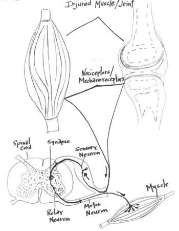
Joints are blessed with four types of mechanoreceptors.  We have covered this in many other posts (see here and here).  These mechanoreceptors apprise the central nervous system of the position (proprioception or kinesthesis) of that body part or joint via the dorsal column system or spinocerebellar tracts. Damage to these receptors can result in a mismatch or inaccuracy of information to the central nervous system (CNS). This can often result in further injury or a new compensation pattern. 

Joints have another protective mechanism called arthrogenic inhibition (see diagram above). This protective reflex turns off the muscles which cross the joint. This was described in a few great paper by Iles and Stokes in the late 80’s an early 90’s (vide infra). Not only are the muscles inhibited, but it can also lead to muscle wasting; there does not need to be pain and a small joint effusion can cause the reflex to occur. 

If the muscles are inhibited and cannot provide appropriate afferent (sensory) and efferent (motor) information to the CNS, your brain makes other arrangements to have the movement occur, often recruiting muscles that may not be the best choice for the job. We call this a “compensation” or “compensation pattern”. An example would be that if the glute max is inhibited (a 2 joint muscle, with a larger attachment to the IT band and a smaller to the gluteal tuberosity; it is a hip extender, external rotator and adductor of the thigh), you may use your lumbar erectors (multi joint muscles; extensors and lateral rotators of the lumbar spine) or hamstrings (2 joint muscles; hip extenders, knee flexors, internal and external rotators of the thigh)  to extend the hip on that side, resulting in aberrant mechanics often observable in gait, which may manifest itself as a shortened step length, increased vertical displacement of the pelvis, lateral shift of the pelvis or increase in step height, just to name a few. Keep this up for a while and the new “pattern” becomes ingrained in the CNS and that becomes your new default for that motion.

Now to fix the problem, you not only need to reactivate the muscle, but you need to retrain the activity. Alas, the importance of doing a thorough exam and thorough rehab to fix the problem.

Often times, the fix is much more involved than figuring out what the problem is (or was). Take your time and do a good job. Your clients and patients will appreciate it!

Ivo and Shawn, the gait guys

Young A, Stokes M, Iles JF : Effects of joint pathology on muscle. Clin Orthop Relat Res. 1987 Jun;(219):21-7

Iles JF, Stokes M, Young A.: Reflex actions of knee joint afferents during contraction of the human quadriceps. Clin Physiol. 1990 Sep;10(5):489-500.

image from: http://chiroeco.com/chiro-blog/results-to-referrals/2013/04/03/neurology-based-simplified-musculoskeletal-assessment/

Podcast 80: Muscle Receptors, Building your Gait Brain

This week’s show sponsors: 

www.newbalancechicago.com

www.lemsshoes.com

A. Link to our server: 

http://traffic.libsyn.com/thegaitguys/pod_79f.mp3

Direct Download: 

http://thegaitguys.libsyn.com/podcast-79

B. iTunes link:

https://itunes.apple.com/us/podcast/the-gait-guys-podcast/id559864138

C. Gait Guys online /download store (National Shoe Fit Certification and more !) :

http://store.payloadz.com/results/results.aspx?m=80204

D. other web based Gait Guys lectures:

www.onlinece.com   type in Dr. Waerlop or Dr. Allen,  ”Biomechanics”

______________

Today’s Show notes:

A revolutionary new implant of regenerative cells … . 
The U.S. Military Wants to Inject People’s Brains With Painkilling Nanobots That Could Replace Medicine
Random thoughts on the Symmetry of the Gait Cycle
Human gait is cyclical. For the most part, when one limb is engaged on the ground (stance phase), the other is in swing phase (in walking gait there is a brief period of double limb support), then th…

Random thoughts on the Symmetry of the Gait Cycle

Human gait is cyclical. For the most part, when one limb is engaged on the ground (stance phase), the other is in swing phase (in walking gait there is a brief period of double limb support), then the limbs switch tasks. For us to move cleanly and efficiently one would assume that the best way to do that would be to ensure that both limbs are capable of doing the exact same things, with the same timing, same skill, same endurance and same strength. For this clean seamless motor function, one must assume that there is complete limb symmetry (length, long bone torsion, the same rate and degree of pronation, supination, ankle dorsiflexion, hip internal/external rotation, same strength, power output etc) and one would hope there would be no injuries that had left a remnant mark on one limb thus encouraging a necessary compensation pattern in that limb (and one that would then have to be negotiated with the opposite limb).  For example, when right ankle rocker (dorsiflexion) is impaired, early heel departure will occur and hip extension will be limited. An alteration in right glute function will follow.  One could theorize that the left step length (the length of measure from right heel strike through to left heel strike) would be shortened. This would cause a premature load onto the left limb, and could very well force the left frontal plane to be more engaged than is desirable. This could lead to left core and hip frontal plane weakness and compensation patterns to be generated. To complicate the cyclical scenario, the time usually used to move sagittally will be partially used to move into, and back out of, the left frontal plane. This will necessitate some abbreviations in the left stance phase’s timely mechanical events. Some biomechanical events will have to be abbreviated or sped through and then the right limb will have to adapt to those changes. These are simple gait problems we have talked about over and over again here on the gait guys blog. These compensation patterns will include weaknesses as part of the pattern, and fixing those weaknesses does not address the right ankle rocker problem. Fixing said weaknesses merely encourages the brain to possibly continue to perpetuate necessary tightnesses in other muscles and motor linkages and engrain the compensations further or more complexly.  It is easy to find something weak, it takes a sharp brain to find the sometimes silent sparking event underneath it all. One’s focused task should be, are you able to find the problem in this never ending loop of compensations and find a way to unwrinkle the system one logical piece at a time, or will you just chose to strengthen the wrinkled system and hope that the new strength on top of the compensations is adequate for you our your client ? One should not have to do daily or weekly rehabilitative sessions and homework to negate and alleviate symptoms, this is a far more durable machine than one that needs daily support.  Rather, one that “seems” to need daily supportive homework/rehab is one that likely needs the underlying limitation to be uncovered. However, there are always exceptions. If one has a fixed issue, for example Foot Baller’s Ankle, then regular doses of lower limb anterior compartment work may be necessary to ensure that further ankle dorsiflexion range is not eroded.  

Now, lets add another wrinkle to the system.  What if there were problems before any injuries ?  Meaning, what if there were problems during the timely maturation and suppression of the primitive reflexes ? Or problems in the timely appearance or maturation of postural reflexes? A problem in these areas may very well result in a central or peripheral nervous system malfunction and a representation of such in one’s movement and gait.  But, that is a discussion for another time.

Shawn and Ivo, the gait guys

photo: courtesy of Thomas Michaud, from the excellent textbook, Human Locomotion

tumblr_ne2sg64OZ11qhko2so1_400.gif
tumblr_ne2sg64OZ11qhko2so3_500.jpg
tumblr_ne2sg64OZ11qhko2so2_1280.jpg

More thoughts on stretching

   We get a lot of interest in our posts on stretching. Seems like this is a pretty hot subject and there is a lot of debate as to whether it is injury preventative or not. Are you trying to physically lengthen the muscle or are you trying to merely bring it to its physiological limit?  There’s a big difference in what you need to do to accomplish each of these goals. Lets take a look at each, but 1st we need to understand a little about muscles and muscle physiology.

 Muscles are composed of small individual units called sarcomeres. Inside of these “sarcomeres” there are interdigitating fibers of actin and myosin (proteins) which interact with one another like a ratchet when a muscle contracts.  Sarcomeres can be of various lengths, depending on the muscle, and are linked and together from one end of the muscle to the other. When a muscle contracts concentrically (the muscle shortening while contracting) the ends of the sarcomere (called Z lines or Z discs) are drawn together, shortening the muscle fiber over all (see the picture above).
 
 Signals are sent from the brain (actually the precentral gyrus of the cerebral cortex areas 4, 4s and 6) down the corticospinal tract to the spinal cord to synapse on motor neurons there.  These motor neurons (alpha motor neurons) then travel through peripheral nerves to the muscles to cause them to contract (see picture above).

   The resting length of the muscle is dependent upon two factors:
The physical length of the muscle
2. The “tone” of the muscle in question.

The physical length of the muscle is determined by the length of the sarcomeres and the number of them in the muscle.   The “tone” of the muscle determined by an interplay of neurological factors and the feedback loops between the sensory (afferent) receptors in the muscle (Ia afferents, muscle spindles, Golgi tendon organs etc.), relays in the cerebellum and basal ganglia as well as input from the cerebral cortex.

 If you’re trying to “physically lengthen” a muscle, then you will need to actually add sarcomeres to the muscle. Research shows that in order to do this with static stretching it must be done 20 to 30 minutes per day per muscle.

 If you were trying to “bring a muscle to its physiological limit” there are many stretching methods to accomplish this.  Pick your favorite whether it be a static stretch, contract/ relax, post isometric relaxation etc. and you’ll probably be able to find a paper to support your position.

  Remember with both not to ignore neurological reflexes (see above). Muscle spindle loops are designed to provide feedback to the central nervous system about muscle length and tension. Generally speaking, slow stretch activates the Ia afferent loop which causes causes physiological contraction of the muscle (this is one of the reasons you do not want to do slow, steady stretch on a muscle in spasm). This “contraction” can be fatigued overtime, causing the muscle to be lengthened to it’s physiological limit.  Do this for an extended period of time (20-30 mins per day) and you will physically add sarcomeres to the muscle.

 Next time you are stretching, or you were having a client/patient stretch, think about what it is that you’re actually trying to accomplish  because there is a difference.

We are and remain The Gait Guys.  Bald, good-looking, and above-average intelligence. Spreading gait literacy with each post we publish.

thanks to scienceblogs.com for the corticospinal tract image

Hip muscles and postural control related to ankle function.

Hip exercises boost postural control in individuals with ankle instability
http://lermagazine.com/news/in-the-moment-sports-medicine/hip-exercises-boost-postural-control-in-individuals-with-ankle-instability

-“Four weeks of hip external rotator and abductor strengthening significantly improves postural control in patients with functional ankle instability (FAI) and may be useful for preventing recurrent instability, according to research from Indiana University in Bloom­ington.”

Nothing new here, at least not here on The Gait Guys blog. We have been talking about these kinds of issues for a long time. We  have long discussed the necessary control of the glutes (and their anchoring abdominals) to eccentrically control the loading response during the stance phase of gait, we especially like to discuss the control of the rate of internal rotation (read: eccentric ability of external rotators as a component) of the leg with the glutes. It is why we think it is so important to eccentrically test the glutes and the core stabilizers (all of them !) when the client is table assessed because it is a huge window for us as to what is happening when there is ground interface. Sure one is open chain and the other is closed, but function is necessary in both. 
What this article is again, like others, telling us is that the ability to stack the joints (knee over foot, hip over knee, level stable pelvis over hip) improves postural control, especially when there is a risky environment of ankle functional or anatomical instability. 
And yes, we are talking Cross over gait and frontal plane challenges and faulty patterns here.  Failure to stack the joints usually leads to cross over gait challenges (type in “cross over or cross over gait into our blog SEARCH box). Remember though, you must selectively strengthen the weak muscles and weak motor patterns, if you are not specific you can easily strengthen the neuro-protective tight muscles and their patterns because they have been the only available patterns to your client. If you are not careful, you will help them strategize and compensate deeper, which in itself can lead to injury.  This is a paramount rehab principle, merely activating what appears weak does not mean you are carrying them over to a functional pattern. Just because you can show a change on the table doesn’t mean it carries over to the ground and sport or training. 
Shawn and Ivo, the gait guys
Why does it feel so good to stretch? 
We are sure you have read many articles, some written by us, about the good the bad and the ugly about stretching.  Regardless of how you slice the cake, we think we can all agree that stretching “feels” good. T…

Why does it feel so good to stretch? 

We are sure you have read many articles, some written by us, about the good the bad and the ugly about stretching.  Regardless of how you slice the cake, we think we can all agree that stretching “feels” good. The question of course is “Why?”

Like it or not, it all boils down to neurology. Our good old friends, the Ia afferents are at least partially responsible, along with the tactile receptors, like Pacinian corpuscles, Merkel’s discs, Golgi tendon organs, probably all the joint mechanoreceptors and well as a few free nerve endings. We have some reviews we have written of these found here, and here and here.

What do all of these have in common? Besides being peripheral receptors. They all pass through the thalamus at some point (all sensation EXCEPT smell, pass through the thalamus) and the information all ends up somewhere in the cortex (parietal lobe to tell you where you are stretching, frontal lobe to help you to move things, insular lobe to tell you if it feels good, maybe the temporal lobe so you remember it, and hear all those great pops and noises and possibly the occipital lobe, so you can see what you are stretching.

The basic (VERY basic) pathways are:Peripheral receptor-peripheral nerve-spinal cord-brainstem-thalamus-cortex; we will call this the “conscious” pathway:  and peripheral receptor-peripheral nerve-spinal cord-brainstem-cerebellum- cortex; we will call this the “unconscious” pathway.

Of course, the two BASIC pathways cross paths and communicate with one another, so not only can you “feel” the stretch with the conscious pathway but also know “how much” you are stretching through the unconscious pathway. The emotional component is related through the insular lobe (with relays from the conscious and unconscious pathways along with collaterals from the temporal lobe to compare it with past stretching experiences) to the cingulate gyrus and limbic cortex,  where stretching is “truly appreciated”. 

As we can see, there is an interplay between the different pathways and having “all systems go” for us to truly appreciate stretching from all perspectives; dysfunction in one system (due to a problem, compensation, injury, etc) can ruin the “stretching experience”. 

Hopefully we have stretched your appreciation (and knowledge base) to understand more about the kinesthetic aspect of stretching. We are not telling you to stretch, or not to stretch, merely offering a reason as to why we seem to like it.

The Gait Guys

Podcast 79: Tightness vs. Shortness, Plantar Fascitis & more.

plus, pelvic asymmetry, “wearables” and cognitive choices in movement.

This week’s show sponsors: 

www.newbalancechicago.com

www.lemsshoes.com

A. Link to our server: 

http://traffic.libsyn.com/thegaitguys/pod_79f.mp3

Direct Download: 

http://thegaitguys.libsyn.com/podcast-79

B. iTunes link:

https://itunes.apple.com/us/podcast/the-gait-guys-podcast/id559864138

C. Gait Guys online /download store (National Shoe Fit Certification and more !) :

http://store.payloadz.com/results/results.aspx?m=80204

D. other web based Gait Guys lectures:

www.onlinece.com   type in Dr. Waerlop or Dr. Allen,  ”Biomechanics”

______________

Today’s Show notes:

The Brain and your choices.

http://exploringthemind.com/the-mind/brain-scans-can-reveal-your-decisions-7-seconds-before-you-decide#.VCx0P8ydUK4.facebook


Walking is the superfood of fitness, experts say
 
Hey Guys,
I have pelvis asymmetry and a snapping ankle, can you help me with … . 
 
New research on Plantar Fascitis
 
John from FB
Shortness vs tightness:
What protocol do you recommend for stretching ? I usually do static stretches1x2min. This article has the static stretch group doing 10x30sec. I’d have to set my alarm a half hour earlier! :-)
Some Fat on Flat FeetNormal feet:
more hindfoot dorsiflexion (read ankle rocker)
hindfoot more flexible
no or different compensation, if any
Symptomatic Flat feet:
less hindfoot dorsiflexion (read, reduced ankle rocker)
hindfoot was more everted, bu…

Some Fat on Flat Feet

Normal feet:

  • more hindfoot dorsiflexion (read ankle rocker)
  • hindfoot more flexible
  • no or different compensation, if any


Symptomatic Flat feet:

  • less hindfoot dorsiflexion (read, reduced ankle rocker)
  • hindfoot was more everted, but less flexible.
  • forefoot compensates for reduced motion in rearfoot by increasing motion 
  • hallux hypermobility
  • symptomatic flat feet lacked positive joint energy for propulsion 


Asymptomatic flat feet:

  • less hindfoot dorsiflexion (read, reduced ankle rocker)
  • hindfoot was more everted, but less flexible.
  • forefoot compensates for reduced motion in rearfoot by increasing motion 
  • hallux hypermobility
  • asymptomatic flat feet needed to absorb more negative ankle joint energy during loading response. This may risk fatigue and overuse syndrome of anterior shank muscles


“Hence, despite a lack of symptoms flatfoot deformity in asymptomatic flat feet affected function. Yet, contrary to what was expected, symptomatic flat feet did not show greater deviations in 3D foot kinematics than asymptomatic. Symptoms may rather depend on tissue wear and subjective pain thresholds.”


http://www.ncbi.nlm.nih.gov/pubmed/23796513

Podcast 78: Step Width Gait, Training Asymmetries & more

Show sponsors: 

www.newbalancechicago.com

www.lemsshoes.com

A. Link to our server: 

http://traffic.libsyn.com/thegaitguys/pod_78ff.mp3

Direct Download: 

http://thegaitguys.libsyn.com/podcast-78

B. iTunes link:

https://itunes.apple.com/us/podcast/the-gait-guys-podcast/id559864138

C. Gait Guys online /download store (National Shoe Fit Certification and more !) :

http://store.payloadz.com/results/results.aspx?m=80204

D. other web based Gait Guys lectures:

www.onlinece.com   type in Dr. Waerlop or Dr. Allen,  ”Biomechanics”

______________

Today’s Show notes:

24-year-old woman missing entire cerebellum exemplifies the amazing power of brain plasticity

Brain scans reveal ‘gray matter’ differences in media multitaskers

Who are we: Ivo talk a bit about yourself and your educational history and what is your website ?
Shawn…..do the same
and……lets keep each interesting but to just a few minutes
Effect of step width manipulation on tibial stress during running
Does Limited Internal Femoral Rotation Increase Peak Anterior Cruciate Ligament Strain During a Simulated Pivot Landing?
http://ajs.sagepub.com/content/early/2014/09/22/0363546514549446.abstract
Quadriceps Muscle Function After Exercise in Men and Women With a History of Anterior Cruciate Ligament Reconstruction
http://natajournals.com/doi/abs/10.4085/1062-6050-49.3.46
tumblr_nd32fvOzjx1qhko2so3_r1_1280.png
tumblr_nd32fvOzjx1qhko2so2_1280.png
tumblr_nd32fvOzjx1qhko2so1_1280.png
tumblr_nd32fvOzjx1qhko2so4_r1_500.png

Part 2: “Standing on Glass” Static Foot/Pedograph Assessment

* note (see warning at bottom): This is a static assessment dialogue. One cannot, and must not, make clinical decisions from a static assessment. The right and left sides are indicated by the R and L circled in pink. There are 4 photos here today.

Blue lines: Last time we evaluated possible ideas on the ORANGE lines here, it would be to your advantage to start there. 

We can see a few noteworthy things here in these photos. We have contrast-adjusted the photo so the pressure areas (BLUE) are more clearly noted. There appears to be more forefoot pressure on the right foot (the right foot is on the readers left), and more rearfoot pressure on the left (not only compare the whiteness factor but look at the displacement of the calcaneal fat pad (pink brackets). There is also noticeably more lateral forefoot pressure on the left. There is also more 3-5 hammering/flexion dominance pressure on the left.  The metatarsal fat pad positioning (LIME DOTS represent the distal boundary) is intimately tied in with the proper lumbrical muscle function  (link) and migrates forward toward the toes when the flexors/extensors and lumbricals are imbalanced. We can see this fat pad shift here (LIME DOTS). The 3-5 toes are clearly hammering via flexor dominance (LIME ARROWS), this is easily noted by visual absence of the toe shafts, we only see the toe pads. Now if you remember your anatomy, the long flexors of the toes (FDL) come across the foot at an angle (see photo). It is a major function of the lateral head of the Quadratus plantae (LQP) to reorient the pull of those lesser toe flexors to pull more towards the heel rather than on an angle. One can see that in the pressure photos that this muscle may be suspicious of weakness because the toes are crammed together and moving towards the big toe because of the change in FDL pull vector (YELLOW LINES). They are especially crowding out the 2nd toe as one can see, but this can also be from weakness in the big toe, a topic for another time. One can easily see that these component weaknesses have allowed the metatarsal fat pad to migrate forward. All of this, plus the lateral shift weight bearing has widened the forefoot on the left, go ahead, measure it. So, is this person merely weight bearing laterally because they are supinating ? Well, if you read yesterday’s blog post we postulated thoughts on this foot possibly being the pronated one because of its increased heel-toe and heel-ball length. So which is it ? A pronated yet lateral weight bearing foot  or a normal foot with more lateral weight bearing because of the local foot weaknesses we just discussed ? Or is it something else ? Is the problem higher up, meaning, are they left lateral weight bearing shift because of a left drifted pelvis from weak glute medius/abdominal obliques ?  Only a competent clinical examination will enlighten us.

Is the compensation top-down or bottom up, or both in a feedback cycle trying to find sufficient stability and mobility ? These are all viable possibilities and you must have these things flowing freely through your head during the clinical examination as you rule in/rule out your hands-on findings.  Remember, just going by a screen to drive prescription exercises from what you see on the movement screen is not going to necessarily fix the problem, it could in fact lead one to drive a deeper compensation pattern. 

Remember this critical fact.  After an injury or a long standing problem, muscles and motor patterns jobs are to stabilize and manage loads (stability and mobility) for adequate and necessary movement. Injuries leave a mark on the system as a whole because adaptation was necessary during the initial healing phase. This usually spills over during the early movement re-introduction phase, particularly if movement is reintroduced too early or too aggressively.  Plasticity is the culprit. Just because the injury has come and gone does not mean that new patterns of skill, endurance, strength (S.E.S -our favorite mnemonic), stability and mobility were not subsequently built onto the apparently trivial remnants of the injury.  There is nothing trivial if it is abnormal. The forces must, and will, play out somewhere in the body and this is often where pain or injury occurs but it is rarely where the underlying problem lives.

Come back tomorrow.  We will try to bring this whole thing together, but remember, it will just be a theory for without an exam one cannot prove which issues are true culprits and which are compensations. Remember, what you see is often the compensatory illusion, it is the person moving with the parts that are working and compensating not the parts that are on vacation.  See you tomorrow friends !

Shawn and ivo, the gait guys

* note: This is a static assessment dialogue. One cannot, and must not, make clinical decisions from a static assessment. As in all assessments, information is taken in, digested and then MUST be confirmed, denied and/or at the very least, folded into a functional and clinically relevant assessment of the client before the findings are accepted, dismissed and acted upon. As we always say, a gait analysis or static pedograph-type assessment (standing force plate) is never enough to make decisions on treatment to resolve problems and injuries. What is seen and represented on either are the client’s strategies around clinical problems or compensations.  Today’s photo and blog post are an exercise in critical clinical thinking to get the juices flowing and to get the observer thinking about the client’s presentation and to help open up the field to questions the observer should be entertaining.  The big questions should be, “why do i see this, what could be causing these observances ?”right foot supinated ? or more rear and lateral foot……avoiding pronation ?

The &ldquo;Standing on Glass&rdquo; Static Foot/Pedograph Assessment: Part 1
* note: This is a static assessment dialogue. One cannot, and must not, make clinical decisions from a static assessment. As in all assessments, information is taken in, di…

The “Standing on Glass” Static Foot/Pedograph Assessment: Part 1

* note: This is a static assessment dialogue. One cannot, and must not, make clinical decisions from a static assessment. As in all assessments, information is taken in, digested and them MUST be confirmed, denied and/or at the very least, folded into a functional and clinically relevant assessment of the client before the findings are accepted, dismissed and acted upon. As we always say, a gait analysis or pedograph-type assessment is never enough to make decisions on treatment to resolve problems and injuries. What is seen and represented on either are the client’s strategies around clinical problems or compensations.  Today’s photo and blog post are an exercise in critical clinical thinking to get the juices flowing and to get the observer thinking about the client’s presentation and to help open up the field to questions the observer should be entertaining.  The big questions should be, “why do i see this, what could be causing these observances ?”

* note the right and left sides by the R and L circled in pink.

ORANGE lines: The right foot appears to be shorter, or is it that the left is longer (see the lines and arrows drawing your attention to these differences)? A shorter foot could be represented by a supinated foot (if you raise the arch via the windlass mechanism you will shorten the foot distance between the rear and forefoot). A longer foot could be represented by a more pronated foot.  Is that what we have here ? There is no way to know, this is a static presentation of a client standing on glass. What we should remember is that the goal is always to get the pelvis square and level.  If an anatomically or functionally short leg is present, the short leg side MAY supinate to raise the mortise and somewhat lengthen the leg.  In that same client, they may try to meet the process part way by pronating the other foot to functionally “shorten” that leg.  Is that what is happening here ? So, does this client have a shorter right leg ? Longer left ?  Do you see a plunking down heavily onto the right foot in gait ? Remember, what you see is their compensation.  Perhaps the right foot is supinating, and thus working harder at the bottom end of the limb (via more supination), to make up for a weak right glute failing to eccentrically control the internal spin of the leg during stance phase ? OR, perhaps the left foot is pronating more to drive more internal rotation on the left limb because there is a restricted left internal hip rotation from the top ? Is the compensation top-down or bottom up ? These are all viable possibilities and you must have these things flowing freely through your head during the clinical examination as you rule in/rule out your hands-on findings.  Remember, just going by a FMS-type screen to drive prescription exercises from what you see on a movement screen is not going to necessarily fix the problem, it could in fact lead one to drive a deeper compensation pattern. You can be sure that Gray Cook’s turbo charged brain is juggling all of these issues (and more !) when he sees a screen impairment, although we are not speaking for him here.

Remember this critical fact.  After an injury or a long standing problem, muscles and motor patterns jobs are to stabilize and manage loads (stability and mobility) for adequate and necessary movement. Injuries leave a mark on the system as a whole because adaptation was necessary during the initial healing phase. This usually spills over during the early movement re-introduction phase, particularly if movement is reintroduced too early or too aggressively.  Plasticity is the culprit. Just because the injury has come and gone does not mean that new patterns of skill, endurance, strength (S.E.S -our favorite mnemonic), stability and mobility were not subsequently built onto the apparently trivial remnants of the injury.  There is nothing trivial if it is abnormal. The forces must, and will, play out somewhere in the body and this is often where pain or injury occurs but it is rarely where the underlying problem lives.

Come back tomorrow, where we will open your mind into the yellow, pink, blue and lime markings on the photo. Are the hammering toes (lime) on the left a clue ? How about the width of the feet (yellow) ? The posturing differences of the 5th toe to the lateral foot border ?  What about the static plantar pressure differences from side to side (blue)? Maybe, just maybe, we can bring a logical clinical assumption together and then a few clinical exam methods to confirm or dis-confirm our working diagnostic assumption.  See you tomorrow friends !

Shawn and ivo, the gait guys

tumblr_nc9gscGC521qhko2so2_1280.jpg
tumblr_nc9gscGC521qhko2so1_1280.jpg
tumblr_nc9gscGC521qhko2so3_1280.jpg
tumblr_nc9gscGC521qhko2so4_1280.jpg
tumblr_nc9gscGC521qhko2so5_1280.jpg
tumblr_nc9gscGC521qhko2so6_1280.jpg

Whoa!  It is amazing what the human frame can withstand…

This 300 pound individual is retired from working with tow trucks from a towing company as well as a service station.   He believes working with the tow trucks, particularly jumping out of them contributed to the O.A. of the ankles.

He has osteoarthritic ankles, a rear foot varus of 15 degrees left side, 5 degrees right.  He is currently in the New Balance 1040 shoe.  He would like some new orthotics built. He Fowler tests positive on his current orthotic set up (with the foot on the ground, dorsiflex the foot at the 1st metatarsal phalangeal joint (ie big toe joint), simulating terminal stance; the orthotic should hug the arch through the range of motion; ie about 45-60 degress of great toe dorsiflexion, which he incredibly has). He is unable to one leg stand because of the O.A. on the ankles and pain.

He has bi-lat. internal tibial torsion, Left > Right and moderate tibial varum, L > R. He has very little internal rotation of the hips bi-lat. Ankle dorsiflexion is about 5 degrees bilaterally.

He is currently in an older New Balance motion control shoe. You can see how he has worn the shoes into varus. More neutral shoes hurt his feet; attempts to put his rear foot into valgus causes increased ankle pain. Exercise compliance is minimal.

WHAT WOULD YOU DO?

The Gait Guys. Teaching and educating with each post.

Shoe lacing problems, things you need to know (that you don't).

How you lace your shoes does truly matter (according to this study).
We have talked about shoe lacing on more than one occasion. Everyone has played around with different laces and lacing strategies at one time or another. And, every shoe seems to lace just a little differently. Some shoes lace far into the forefoot, some have the potential to lace high up into the ankle.  But just because there are eyelets there, doesn’t mean  you have to thread a lace through the hole. It is about fit the majority of the time.  Some of our runners will use “skip” lacing to avoid pressure over the dorsum of the foot, especially if they have a saddle exostosis or hot tendon in that area, much of the time this works to alleviate the pain and pressure there. Just remember, impaired ankle rocker often via weakness of the anterior compartment muscles (toe extensors, tibialis anterior, peroneus tertius) will force dorsiflexion moments into the midfoot and can cause some joint-related compressive pressure on the dorsal foot which can seemingly (and mistakenly) come from shoes tied too tight across the top of the foot. Be sure to consider this fact before you “skip lace” your shoes, it is a big player, one we see all of the time.
In today’s journal article found below, we discover some other factors in a controlled study.  Here they look into the effects of lacing on biomechanics in running, specifically rearfoot runners. The results of the study showed reduced loading rates and pronation velocities as well as lowest peak pressures under the heel and lateral midfoot in the tightest and highest laced shoes. Whereas, the lower laced shoes resulted in lower impacts and lower peak pressures under the 3rd and 4th metatarsal heads (they proposed that this was from forward foot slide in the shoe because of this lacing). The study authors concluded 

 A firm foot-to-shoe coupling with higher lacing leads to a more effective use of running shoe features and is likely to reduce the risk of lower limb injury.

Remember, this is just data for you to cogitate over. It can help you work through some possible issues with your feet and your sport, however it does not translate to everyone as a standard protocol. Remember this, we have been known to say, “your problem is not often the shoe, it is the thing in the shoe (you and your faulty biomechanics)”. However, blaming your problems on you is not good shoe manufacturer advertising, so many shoe companies will offer a plethora of shoes choices for you to accommodate to your variables. This does not necessarily mean the problem is solved, rather it is often managed by a “better” shoe choice that seems to work for your variables. This is a good thing most of the time, if you understand shoes, shoe anatomy, and human anatomy (foot types) so that you can pair them up for a best outcome. The problem may lie in the fact that your shoe fitter is not likely to have all of the necessary pieces to put your perfectly matched picture together, including understanding your total body biomechanics and possibly understanding why a weak glute is impairing hip extension and thus limiting ankle rocker motion, causing premature heel rise, and thus forcing too much dorsiflexion into the arch of the foot and premature forefoot loading causing what seems to be too tightly tied shoes.  
What we truly need an e-Harmony for matching shoes and feet ! But since that perfect scenario doesn’t often exist at the shoe store level or gait analysis level, here at The Gait Guys we have put together the next best thing, The National Shoe Fit Certification Program if you care to take this all to the next level. 
Shoe fitting is an art, and lacing is just another paint brush you can  use to get the job done. You just have to know what brush to use for each given piece of art (ie. the athlete). 
Shawn and Ivo, The gait guys
J Sports Sci. 2009 Feb 1;27(3):267-75. doi: 10.1080/02640410802482425.

Effects of different shoe-lacing patterns on the biomechanics of running shoes.


Does this guy have a short leg or what? How good are your eyes?

One again, we had the gait cam, investigating gait on the east coast. What do we see in this gent?

  • heel strike on out side of left foot with increased progression angle

he appears to be stabilizing the left side during stance phase. notice the upper torso shift to the left during left stance phase

  • abbreviated arm swing on right

note that ankle rocker is adequate on the left

  • body lean to right on right stance phase

gluteus medius weakness on right? short leg on right?

Good.

  • Did you also notice the loss of ankle rocker on the right, compared to the left? This results in less hip extension on that side as well.
  • He flexes his right thigh less than his right during pre swing and swing

external obliques should be firing to initiate hip flexion, perpetuated by the psoas, iliacus and rectus femoris. This does not appear to be happening.

All of this is great BUT nothing like being able to actually examine your patients is there? You can see how gait analysis can tell us many things, but they need to be confirmed by a physical exam.

The Gait Guys. Educating (and hopefully enlightening) with each post. Keep your eyes open and your thinking from the ground up : )


Got Arm Swing?

We have written many times about arm swing. Click here for some of our posts here on Tumblr.

Here we are again at the beach. Look at the beautiful difference in arm swing from side to side in the guy carrying the bag. Makes you want to tell him to use a backpack, eh?

Never mind what it does to his gait

  • decreased arm swing on the carrying side
  • increased step length on the left side
  • increased thigh flexion of the left side
  • increased body lean and head tilt to right side (Take a look at this paper)

think about the increased metabolic cost. Think about what this  type of input (increased amplitude of movement unilaterally) is doing to your cortex!

keep your movements symmetrical, folks!

The Gait Guys

A profound loss of hip extension…

While sitting on the beach, our mind never rests. Even when on vacation we continue to watch how people move.

Luckily today, I had the gait cam (Dr Allen is holding down the Gait Guys Fort), so live from Sunset Beach, it’s Sunday night. See of you can see what I saw.

Sitting with my wife and watching the kids dig in the sand, this gal with the flexed posture caught my eye.

Why is she so flexed forward? The profound loss of hip extension made it impossible for her to stand up straight! It was difficult to say if she has bilateral hip osteoarthritis, or possible bilateral THR’s (total hip replacements), maybe just really tight hip flexors, painful bunions that do not like toe off, or even all of the above. She may have a leg length discrepancy, as she leans to the left on left stance phase; of course she could have weak hip abductors on the left. It does not appear she has good control of her core.

What do we see?

  • flexion at the waist
  • loss of hip extension
  • body lean to left at left midstance
  • shortened step length
  • loss of ankle rocker
  • premature heel rise
  • decreased arm swing (she is carrying something in her left hand)

No one is safe from the gait cam! Stay tuned for more beach footage this week!

We remain, The Gait Guys, even on vacation.