tumblr_m0lqunwlic1qhko2so1_1280.jpg
tumblr_m0lqunwlic1qhko2so2_1280.jpg

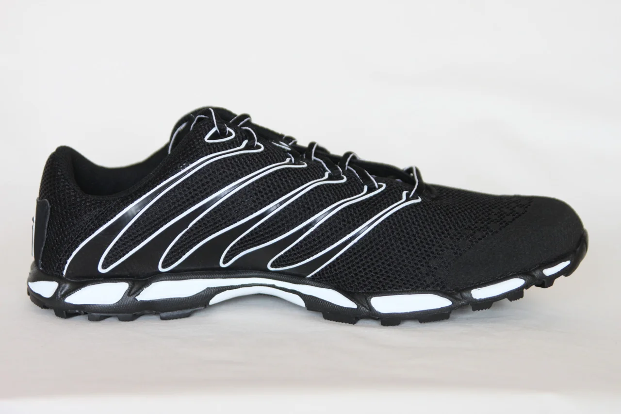
The Sole Truth and Nothing but the Truth

Thicker soles mean more muscle activity. Nothing new here. We have posted on the fallacy of increased cushioning and decreased impact many times. Here is another supporting study.


Here are part of the results:Compared to the barefoot condition, there is an increase in the magnitude of muscle contraction on wearing shoes, which further increases with thickening shoe soles.”

and the conclusion...“Footwear with increasing shoe sole thickness evokes a correspondingly stronger protective eversion response from the peroneus longus to counter the increasing moment at the ankle-subtalar joint complex following sudden foot inversion. Hence, fashion footwear with thicker sole is likely to increase the risk of lateral ligament injury of the ankle when such protective response is overwhelmed. Similarly, the clinicians need to be cautious regarding the amount of shoe raise that they could provide for patients with limb length discrepancy without any detrimental untoward side effects.”


We remember the peroneus longus attaches from the upper, lateral fibula, traveling down the fibular shaft, around the lateral malleolus and attaching to the base of the 1st  1st metatarsal and lateral cunieform. It fires from just prior to heel strike to terminal stance, assisting in eversion of the foot and cuboid,  locking the lateral column of the foot during supination, and plantar flexes the 1st ray (brings the medial tripod down to the ground). More sole = More activity = More potential for injury

more on peroneus here


The Gait Guys. Bringing you the science of shoes and the impact on gait, every day.


http://www.ncbi.nlm.nih.gov/pubmed/22017890

Foot Ankle Surg. 2011 Dec;17(4):218-23. Epub 2010 Sep 17.

The influence of shoe sole’s varying thickness on lower limb muscle activity.

Source

Institute of Motion Analysis & Research, Department of Orthopaedic & Trauma Surgery, TORT Centre, Ninewells Hospital & Medical School, University of Dundee, Dundee, DD1 9SY, Scotland, UK.

Abstract

BACKGROUND:

The lateral ligament injury of the ankle is acknowledged to be the most common ankle injury sustained in sport. Increased peroneus longus muscle contraction in the shod population has already been documented. This study aimed to quantify the effect of shoe sole’s varying thickness on peroneus longus muscle activity.

METHODS:

Electromyographic recordings of the peroneus longus muscle activity following unanticipated inversion of the foot from 0° to 20° in a two-footplate tilting platform were collected from 38 healthy participants. The four test conditions were: barefoot, standard shoe, and shoes with 2.5 cm and 5 cm sole adaptation respectively.

RESULTS:

Compared to the barefoot condition, there is an increase in the magnitude of muscle contraction on wearing shoes, which further increases with thickening shoe soles. The peroneus longus was responding earlier in the shod conditions when compared to the barefoot, although the results were variable within the three shod conditions.

CONCLUSION:

Footwear with increasing shoe sole thickness evokes a correspondingly stronger protective eversion response from the peroneus longus to counter the increasing moment at the ankle-subtalar joint complex following sudden foot inversion. Hence, fashion footwear with thicker sole is likely to increase the risk of lateral ligament injury of the ankle when such protective response is overwhelmed. Similarly, the clinicians need to be cautious regarding the amount of shoe raise that they could provide for patients with limb length discrepancy without any detrimental untoward side effects.

Copyright © 2010. Published by Elsevier Ltd.

The Confusion of Shoe Science (Part 3 of 5): Narrowing the Gap Between Science, Manufacturer, Retail and Consumer

* note, if you rushed ahead and watched PART 3 on the IRRC youtube page,  you will now want to watch this critiqued version by Blaise Dubois. This is important that you watch this version so you can get a clearer truth and less of a commerical bias, in our opinion.

This is an important series of lectures / debate.  This event occurred on December 6th, 2011 at The Running Event which we also lectured at.  The result of this series of talks, which we will post 2-5 sequentially daily here on our blog, was an underground heated dialogue between some of the speakers. 

The Gait Guys have no vested interest with any company other than the truth. In our opinion Simon Bartold’s lecture here is very biased towards the company he works for, Asics.

Watch these videos and learn, and make your own conclusions.  But, if you are a runner or shoe store owner or staff, you need to watch these and open your mind and consider that what you are being told by your shoe vendors might not always be the truth.  Educate yourself, ask the hard questions of your shoe vendors and companies and make your own conclusions. 

* If you wish to watch this version without the important additions added by Blaise, you can go here and which Simon’s commercially biased lecture unabridged. click here

Shawn and Ivo  ……. bringing this stuff to you so you do not have to find it on your own.

The Confusion of Shoe Science (Part 2 of 5): Narrowing the Gap Between Science, Manufacturer, Retail and Consumer

This is an important series of lectures / debate.  This event occurred on December 6th, 2011 at The Running Event which we also lectured at.  The result of this series of talks, which we will post 2-5 sequentially daily here on our blog, was an underground heated dialogue between some of the speakers. 

We have come to greatly respect Blaise Dubois for his vast wealth of knowledge and his unbiased standpoint. Blaise is a physicial therapist with worldwide respect for his knowledge and experience.  Unlike some of the other speakers you will hear in the coming days, he had no vested interest other than sharing the facts and myths in this field.  This is why we have enjoyed our growing relationship with Blaise, because we too have no vested interest with any company other than the truth. 

Watch these videos and learn, and make your own conclusions.  But, if you are a runner or shoe store owner or staff, you need to watch these and open your mind and consider that what you are being told by your shoe vendors might not always be the truth.  Educate yourself, ask the hard questions of your shoe vendors and companies and make your own conclusions. 

Shawn and Ivo  ……. bringing this stuff to you so you do not have to find it on your own.

tumblr_m0jshuyehE1qhko2so1_500.jpg
tumblr_m0jshuyehE1qhko2so2_500.jpg
tumblr_m0jshuyehE1qhko2so3_1280.jpg

Boots make the Man (or the Woman)

If you had any doubts about shoes changing ground reaction forces (GRF’s), then read no further.GRF’s are one of the factors considered by some sources as being injurious.

Tennis shoes had the most ground reaction forces

Army boots came in a narrow second

Bare feet had the least forces by a significant amount

Incidently, the amount of the valgus in the knees on landing was not significantly different among the 3 cases.

Hmmm. We need to think before recommending a shoe.

Ivo and Shawn. The Gait Guys. Sifting through the literature so you don’t have to. Yes, we are still bald, middle aged and good looking….

J R Army Med Corps. 2011 Sep;157(3):218-21.

A kinematic and kinetic analysis of drop landings in military boots.

Abstract

OBJECTIVE:

The purpose of this study was to examine knee valgus in drop landings during three different footwear conditions and to examine the ground reaction forces exhibited during the drop landing in the three different footwear conditions.

METHODS:

Sixteen male and female Reserve Officer Training Corps (ROTC) university undergraduate cadets (21 +/- 3 yrs, 79 +/- 12 kg, and 172 +/- 10 cm) volunteered to participate in the study. Kinematic data were collected while participants performed drop landings in three conditions: bare feet, tennis shoes, and issued military boots.

RESULTS:

Significant differences among footwear for ground reaction forces (bare feet: 1646 +/- 359%, tennis shoe: 1880 +/- 379%, boot: 1833 +/- 438%; p < 0.05) were found, while there was no significant difference in knee valgus among footwear.

CONCLUSIONS:

Though footwear conditions did not affect knee valgus, they did affect ground reaction forces. Participants in this study had yet to receive any military training on how to land properly from a specified height. Further research should be completed to analyze the kinematics and kinetics of the lower extremity during different landing strategies implemented by trained military personnel in order to better understand injury mechanisms of drop landings in this population. It is likely that injury prevention landing techniques would be beneficial if these were employed by the military and not just in the sporting community.

tumblr_m0jrg50UgG1qhko2so1_1280.gif
tumblr_m0jrg50UgG1qhko2so2_1280.jpg

Shoe News You Can Use

Pronation is dorsiflexion, eversion and abduction, It is a term usually used to refer to midfoot motion. It occurs as the friction of the heel with the ground causes the talus to slide anteriorly on the calcaneus, as it does so, it plantar flexes, adducts and everts. this motion causes a medial spin of the talus, which, in turn, causes an internal spin of the tibia (see above).

Pronation is a natural motion which  is one of the 4 shock absorbing mechanisms we have to attenuate force (pronation, ankle dorsiflexion,knee flexion and thigh flexion). Some pronation is necessary for normal gait. Over or under pronation appear linked to increased likelihood of injury. Of the 2, over pronation is the most treated (possibly wrongly so) and one intervention is motion control shoes.

Motion control shoes usually have a feature (medial posting, varus positioning, dual density midsoles, increased lateral flare, etc) which attenuates or delays pronation.

Along those lines, an in light of our latest series of posts, we thought you may find this study interesting. Results were as follows: “A one-tailed paired t-test indicated a statistically significant decrease in the total range of proximal tibial rotation when a motion control shoe was worn (mean difference 1.38°, 95% confidence interval 0.03 to 2.73, P=0.04).”

So, motion control shoes decreased motion about 1-1.5 degrees. The average amount of midfoot motion is 4-8 degrees. Our question to you is “Is that enough, or is that significant?”

We think so, especially in some cases. A few degrees can make all the difference. There appears a time and place for motion control shoes, but on our opinion, they are grossly over prescribed for problems that are usually able to be treated more conservatively.

The Gait Guys. Promoting Gait and Foot literacy…everywhere

Physiotherapy. 2011 Sep;97(3):250-5. Epub 2011 Feb 2.

Effect of motion control running shoes compared with neutral shoes on tibial rotation during running.

Abstract

OBJECTIVE: To determine whether a motion control running shoe reduces tibial rotation in the transverse plane during treadmill running.

DESIGN: An experimental study measuring tibial rotation in volunteer participants using a repeated measures design.

SETTING: Human Movement Laboratory, School of Health Professions, University of Brighton.

PARTICIPANTS: Twenty-four healthy participants were tested. The group comprised males and females with size 6, 7, 9 and 11 feet. The age range for participants was 19 to 31 years.

MAIN OUTCOME MEASURES: The total range of proximal tibial rotation was measured using the Codamotion 3-D Movement Analysis System.

RESULTS: A one-tailed paired t-test indicated a statistically significant decrease in the total range of proximal tibial rotation when a motion control shoe was worn (mean difference 1.38°, 95% confidence interval 0.03 to 2.73, P=0.04).

CONCLUSIONS: There is a difference in tibial rotation in the transverse plane between a motion control running shoe and a neutral running shoe. The results from this study have implications for the use of supportive running shoes as a form of injury prevention.

Copyright © 2010 Chartered Society of Physiotherapy. Published by Elsevier Ltd. All rights reserved.

The Confusion of Shoe Science (Part 1 of 5): Narrowing the Gap Between Science, Manufacturer, Retail and Consumer

This is an important series of lectures / debate.  This event occurred on December 6th, 2011 at The Running Event which we also lectured at.  The result of this series of talks, which we will post 2-5 sequentially daily here on our blog, was an underground heated dialogue between some of the speakers. 

We have come to greatly respect Blaise Dubois for his vast wealth of knowledge and his unbiased standpoint. Blaise is a physicial therapist with worldwide respect for his knowledge and experience.  Unlike some of the other speakers you will hear in the coming days, he had no vested interest other than sharing the facts and myths in this field.  This is why we have enjoyed our growing relationship with Blaise, because we too have no vested interest with any company other than the truth. 

Watch these videos and learn, and make your own conclusions.  But, if you are a runner or shoe store owner or staff, you need to watch these and open your mind and consider that what you are being told by your shoe vendors might not always be the truth.  Educate yourself, ask the hard questions of your shoe vendors and companies and make your own conclusions. 

Shawn and Ivo  ……. bringing this stuff to you so you do not have to find it on your own.

Neuromechanics Weekly: Pain Matters


We know that joint swelling (and thus inflammation) inhibits the contraction of the muscle which crosses the joint from the landmark work of Iles and Stokes back in the late 80&rsquo;s. Now here is a paper stati…
Neuromechanics Weekly: Pain Matters


We know that joint swelling (and thus inflammation) inhibits the contraction of the muscle which crosses the joint from the landmark work of Iles and Stokes back in the late 80’s. Now here is a paper stating that pain does the same thing


This tells us that there is an axon collateral from the primary pain neuron (the “C” fiber) that somehow inhibits the alpha moto neuron, similar to a flexor reflex, as pictured. his is most likely through affecting the gamma moto neuron (which goes to the spindles) rather than the alpha motoneuron; so the “sensitivity” of the muscle is changed (remember, spindles detect length changes, golgi’s tension).


So what does this mean to us and gait? It tells us that pain will inhibit the activity (voluntary and involuntary) of the ability for one to use their muscles, especially those crossing the joint in questions. Be aware of inflammation (painful or non painful) or the painful joint, which can contribute to a compensation pattern.


Ivo and Shawn…The Gait Guys. Making your life less painful and more functional….


Muscle Nerve. 2000 Aug;23(8):1219-26.

Inhibition of motor unit firing during experimental muscle pain in humans.

Source

Center for Sensory-Motor Interaction, Orofacial Pain Laboratory, Aalborg University, Denmark.

Abstract

Electromyographic activity was recorded in the masseter muscle to investigate whether the firing characteristics of single motor units (SMUs) were affected by muscle pain. Capsaicin was injected into the masseter to induce pain. The interspike interval (ISI) and recruitment threshold of SMUs were measured while subjects performed isometric contractions at 5, 7.5, 10, 15, and 20% of maximum voluntary contraction. All subjects were able to maintain a stable isometric force during pain, but the mean ISI was significantly increased without changing the recruitment threshold. In all the experimental conditions, the firing frequency increased with increasing force, and SMUs recruited at low force fired at higher rates for all force levels. These results suggest that acute stimulation of nociceptive muscle afferents inhibits SMU activity without changing recruitment order in the homonymous muscle.

Copyright 2000 John Wiley & Sons, Inc.

Sorry for the late post, folks&hellip;We both had pretty crazy days at our clinics&hellip;
Yup, you saw it here 1st. We couldn&rsquo;t believe it either. Look what we found in the Harvard archives.
Sport féminin : saut sur échasses. Korean women on …

Sorry for the late post, folks…We both had pretty crazy days at our clinics…

Yup, you saw it here 1st. We couldn’t believe it either. Look what we found in the Harvard archives.

Sport féminin : saut sur échasses. Korean women on stilts jumping hurdles in a chase, for fun. Advert for Liebig’s Extract of Meat Company. 1904. Via Harvard U.

What does meat extract have to do with women on stilts, jumping hurdles? We could use some help on this one. Anyone have any suggestions?

The Gait Guys. Not on stilts, but teetering while jumping hurdles sometimes….

A case of severe mechanical gait challenges.

This is a unique case. This is a complicated case, there is so much going on. If your eye is getting good at this gait analysis stuff you will know that just from the first pass this gait is very troubled.

This young middle distance runner who came to see us with complaints of chronic anterior and posterior shin splints. This is unusual because usually only one of the lower limb compartments are strained, either the anterior (tibialis anterior mostly) or the posterior compartment (tibialis posterior mostly). Admittedly this is not a fast runner but they love to run none the less, so you do what you can to help.

Please watch this video again and note the following:

  1. crossing over of the knees at the midline (this indicates a scissored gait / circumductory motion of the limbs)
  2. slightly wider based gait compared to knee postioning but neutral compared to hip spacing
  3. client starts heavily on the outer edge of the feet and moves medially
  4. client over strides (step length is increased) which is particularly evident when they are walking towards the camera
  5. early bunion formation and troubles engaging the big toe during stance phase
  6. the knees / patella also appear medially positioned in an environment of a neutral foot progression angle
  7. if you look carefully you can see that they rear foot immediately moves into a valgus posturing (this is rearfoot pronation) and they are also pronating into the forefoot heavily. Interestingly they have decent arch height.

Wow ! So much going on ! This is a gait from hell in some respects. So, what is driving so much of the terrible gait mechanics ? The answer is a congenital loss of ankle rocker (dorsiflexion) bilaterally. This client can barely squat because the ankles just do not dorsiflex. There was clear osseous lock at barely 90 degrees.

Lets break each one down.

  1. Crossing over of the knees at the midline (this indicates a scissored gait / circumductory motion of the limbs). * This is occuring due to some genu valgum of the knees (slightly “knock-knee”). When the knees are valgum they are at risk for brushing together during gait. The client has no choice but to circumduct the limbs to avoid this behavior. Unfortunately they cannot abduct the thighs far enough during many of the gait cycles and so a “Scissored” appearance occurs where the thighs brush and cross over in appearance.
  2. Slightly wider based gait compared to knee positioning but neutral compared to hip spacing. * This is closely related to our answer in #1. Valgus knees will widen the foot spacing side to side because the feet are not under the knee joints. Then couple this with the necessity to circumduct to avoid knees from contacting and the foot posturing is that of an even wider based gait. This can also occur from many hip problems. However as in this case with a congenital loss of ankle rocker, the client uses more foot pronation to progress the tibia over the talus (allowing the tibia to get past 90degrees) and allow them to move forward. This added pronation does magnify and likely progress the knee valgum but there are few other options for this client. This is often a destructive vicious cycle with few good outcomes decades down the road.
  3. Client starts heavily on the outer edge of the feet and moves medially. *This may be to avoid the immediate rear foot pronation that is seen here.
  4. Client over strides (step length is increased) which is particularly evident when they are walking towards the camera. * This may be a conscious attempt to lengthen the shortened stride that occurs because of the limited ankle dorsiflexion ranges. It appears at many moments however to be a result of the extra effort to circumduct the legs sufficiently. A longer stride does play into #3 above, a larger stride usually leads to a heavier lateral heel strike but it also means that the rearfoot pronation will be more aggressive, this is a negative resultant outcome.
  5. Early bunion formation and troubles engaging the big toe during stance phase. *We are not surprised here. Whenever pronation is excessive the first metatarsal (medial foot tripod) is unstable and this changes the mechanics of the hallux muscles to pull towards the 5th metatarsal anchor generating the bunion. Look at the origin and insertion of the adductor hallucis muscle particularly the transverse head, if the 1st MET is anchored the 5th MET is pulled to the 1st and the transverse arch is formed. However, if the 1st MET is unstable and the 5th is the only anchor, the adductor hallucis will pull the toe laterally and form a bunion and hallux valgus and compromise the transverse arch. (particularly look at the left big toe at the :09 to :11 second mark, the big toe and first MET are clearly not anchored to the ground).
  6. The knees / patella also appear medially positioned in an environment of a neutral foot progression angle. * Answers for #1-#5 clearly will medial patellar deviation and drive patellar tracking problems.
  7. If you look carefully you can see that they rear foot immediately moves into a valgus posturing (this is rearfoot pronation) and they are also pronating into the forefoot heavily. Interestingly they have decent arch height, but remember, that does not mean that pronation is not occurring. * This is a result of the loss of ankle rocker mechanics. If they start pronation early at the rear foot it will drive more pronation. When pronation is driven excessively the arch can drop, and with more arch height drop the tibial will pitch forward past the magical 90 degree mark and allow forward motion to occur.

So, how can they run with all this going on ? Well, the answer is quite simple. They avoid most of these issues as best they can. How you ask ? Forefoot strike; they run avoiding heel strike and midfoot strike. By staying on the forefoot all of these rear and midfoot mechanical limitations as well as ankle rocker loss can be avoided by remaining on the forefoot. This makes distance running difficult but anything below the two mile mark is tolerable and the 100-800 distances are probably best suited for their feet. Incidentally they enjoy the 400 the best, no wonder. Also, moving at increased speed will necessitate a forward lean, and a forward lean makes the tibia progression over the talus easier taking out some of the ankle rocker limitations.

This is a foot type, with complications, that is really beyond much of what anyone can do conservatively. We would even argue that surgery is not an option, just a change in activity choice. This is simply a client that should not run beyond distances where they can stay on the forefoot. The foot, ankle and lower limb mechanics just suffer far to much from having to compensate (as discussed in #1-7) to enable pain and problem free running with anything other than forefoot loading. This means that walking is going to be difficult and problematic, as you can see from this video above.

Our only solution in this case ? ……… utilizing a rocker based footwear. Easy Spirit Get UP and Go (link) was our recommendation and it worked very well for this client for walking. Here is a link to this shoe and pictures of the huge forefoot rocker that helps (somewhat) to dampen the mid-forefoot rocker issues but there is not much that can be done for the rear foot rocker issues as discussed. If you use an orthotic to block the rearfoot valgus motion and rearfoot pronation you will pass more challenges to the midfoot-arch and forefoot. Sadly.

This was a very tough case. Getting every aspect of the case in your head during an evaluation is sometimes a challenge. Sometimes you need to see them a 2nd or 3rd time to digest it all. But be patient with yourself, it takes time to get decent at this stuff. This is a perfect case for “getting a feeling and flow” of the persons gait, at their speed. A case evaluation like this on a treadmill or via video analysis can make things tougher because the treadmill can change the dynamics (did you read our Treadmill article in last months Triathlete magazine ? It was linked on the blog 2 weeks ago) and make the client move at its speed and not their speed inhibiting and promoting different mechanics. There are times for a treadmill and times to avoid them. This is an art, in time you will know when to use and when not to use.

Happy Monday Gait Gang………. welcome to The Gaits of Hell !

Shawn and Ivo ……….two gnarly lookin dudes with pitchforks and a toothy grin.

Rolling patterns and their use in body assessment.

First a brief review from yesterday where we talked about the stabilizing function of the diaphragm possibly being an etiological factor in spinal disorders.  Yesterday we included a link to an abstract by the great Dr. P. Kolar.  It considered the correlation between the dynamics of the diaphragm in posture and chronic spinal disorders.  In review of that paper what they found seemed to indicate that poor diaphragm positioning, posturing and control correlated well in their sampling of chronic low back pain clients. The study found smaller diaphragm movements and a higher diaphragm positioning/posturing.  The study found maximum changes in the rib (costal) intervals and middle areas of the diaphragm which asks one to consider the absolute critical importance of thoracic mobility. Extension, lateral flexion and rotation are frequently reduced in chronic back pain clients but we find it rampant in many clients and athletes.  We also find and encourage you to look for, assess, and normalize your clients abdominal oblique, transverse abdominus and rectus abdominus control.  Failure to properly and adequately anchor the lower rib cage to the pelvis via the abdominal wall (the whole wall, circumferentially around the entire torso to the spine) will result in asymmetrical breathing patterns.  And abnormal breathing patterns lead to abnormal spine motion and mobility.

Yesterday we spoke about the need to assess, and if necessary treat, anything that impairs the diaphragm, breathing patterns, thoracic spine mobility and rib cage movement and flexibility.  Rolling patterns as seen above, and here is the Rolling Pattern for Upper Body Drivers (link), are helpful in determining some loss of coordination of the upper or lower body drivers, impaired thoracic spine mobility as well as loss of symmetrical abdominal skill and strength.  Remember, impairment of a primitive movement pattern like rolling is important to be aware of.  The last thing you want to do is drive your training or treatments therapies and rehab efforts into an asymmetrical pattern. These rolling patterns are first developed as a child to learn to turn over. It is a precursor to pressing up the torso like in a push up, which is of course a precursor to crawling, then cruising and then walking.

  Just like yesterday, we come full circle !  From breathing and the diaphragm to rolling and gait…… it is all connected.  Any faulty strategy or pattern driven into the body, even breathing, can impair gait.  And remember, because with gait we have to attach anti-phasic arm swinging with leg swinging. Anything that disturbs this anti-phasic patterning, such as low back pain or loss of thoracic mobility, will drive contralateral arm-leg swing to phasic patterning.

These are nice, simple assessments.  Hope you enjoyed another window into what we do every day when dealing with athletes, patients and runners.  It is all a part of restoring the symmetrical function to a body.

Shawn and Ivo ……. Rock and Rollers.

The Roll of Breathing and Diaphragm Control in Gait, Running and Human Locomotion

In this video you will see many great things. This video of Rickson Gracie is a testament to free fluid movement and body control.  Great athletes do not just practice one thing.  There is some great demonstrations of breathing and diaphragm control at the 3 minute mark, and we will try to parlay this nicely into today’s brief discussion on the Diaphragm.

Abnormal stabilizing function of the diaphragm may be one etiological factor in spinal disorders.  Today we have included a link to an abstract by the great and brilliant Dr. P. Kolar who we have studied under.  It considers the correlation between the dynamics of the diaphragm in posture and chronic spinal disorders.  What they found seemed to indicate that poor diaphragm positioning, posturing and control correlated well in their sampling of chronic low back pain clients. The study found smaller diaphragm movements and a higher diaphragm positioning/posturing.  The study found maximum changes in the rib (costal) intervals and middle areas of the diaphragm which asks one to consider the absolute critical importance of thoracic mobility. Extension, lateral flexion and rotation are frequently reduced in chronic back pain clients but we find it rampant in many clients and athletes.  We also find and encourage you to look for, assess, and normalize your clients abdominal oblique, transverse abdominus and rectus abdominus control.  Failure to properly and adequately anchor the lower rib cage to the pelvis via the abdominal wall (the whole wall, circumferentially around the entire torso to the spine) will result in asymmetrical breathing patterns.  And abnormal breathing patterns lead to abnormal spine motion and mobility. We frequently have to treat and instruct proper breathing patterns to help normalize lateral and posterior rib cage expansion and decent in athletes and clients, particularly those with low back issues but that is not an exclusive group to this problem. Tomorrow we will show you some simple but great videos showing rolling patterns and we will want you to think back to today’s blog post here on how loss of thoracic mobility in extension, rotation and lateral bend as well as loss of symmetrical abdominal skill and strength can impair a primitive movement pattern like rolling. This is a pattern that is first developed as a child to learn to turn over. It is a precursor to pressing up the torso like in a push up, which is of course a precursor to crawling, then cruising and then walking.

See, we were finally able to come full circle !  From breathing and the diaphragm to gait…… it is all connected.  Any faulty strategy or pattern driven into the body, even breathing, can impair gait.  Because with gait we have to attach anti-phasic arm swinging with leg swinging. Anything that disturbs this anti-phasic patterning, such as low back pain, will drive contralateral arm-leg swing to phasic patterning. Don’t think this is important to athletes and humans ? Well, you must have missed our 2 part blog series on Arm Swing.  We provide those links here. Part 1 link and Part 2 link

If you are an athlete, coach, or in the medical movement assessment or gait analysis field……heck, if you study the human body at all and you are not looking at or into arm swing you are not doing what we are doing. And you are missing the bigger boat. So many “gait specialists” and “gait analysis” programs are not even capturing the arm swing let alone looking at it and discovering its critical importance. Did you miss our dialogue on frozen shoulder and impaired contralateral hip dysfunction ?  If you look for it, which many in the therapy world are not, you will see why we treat that opposite lower limb.  Maybe the rest of the folks around the world will catch on in time.  We are slowly getting there, we now have readership in 23 countries, and growing.  If only we had more time, the apocalypse of December 21, 2012 is coming on fast !

The article also found maximal changes in the middle diaphragm areas which suggests looking at the psoas, quadratus lumborum and crus because of their fascial blending into the diaphragm from below.  Thus, investigation of many muscles from below must also be a part of your assessment or training.  But we will save this discussion for another blog post.

We hope you can see that after a year of blog posts (over 500) that you can begin to see the method of our obvious madness.  That being that everything is important for human gait. Remember, we will blend this blog post into the roll assessments you will see on tomorrows post.  So ya’ll come back now……. ya hear ? 

In closing, it is blog posts like this one that we always hope will go viral on the internet. Especially because it has links to two previous articles we wrote on arm swing which we feel are so very important and commonly overlooked.  And we have more arm swing stuff to share, we just need more time.  Consider linking this article to your website, sending it to friends in the fields we discussed. This information is important. It is why we take the time every day to write and share our 40+ years of clinical experience for free. Because the world needs to know this stuff so more people can be helped all over the world.  Consider sharing this with someone or linking it to your Facebook page or website or slap it up on someones forum to create dialogue. Thanks.

The leg bone is connected to the thigh bone…. as the song goes…….

Shawn and Ivo

_________________________________

here is Kolar’s abstract……

J Orthop Sports Phys Ther. 2011 Dec 21. [Epub ahead of print]

Postural Function of the Diaphragm in Persons With and Without Chronic Low Back Pain.

Abstract

OBJECTIVES:To examine the function of the diaphragm during postural limb activities in patients with chronic low back pain and healthy controls.

BACKGROUND: Abnormal stabilizing function of the diaphragm may be one etiological factor in spinal disorders, but a study designed specifically to test the dynamics of the diaphragm in chronic spinal disorders is lacking.

METHODS: Eighteen patients with chronic low back pain due to chronic overloading, ascertained via clinical assessment and MRI examination, and 29 healthy subjects were examined. Both groups presented with normal pulmonary function test results. A dynamic MRI system and specialized spirometric readings with subjects in the supine position were used. Measurements during tidal breathing (TB), isometric flexion of the upper or lower extremities against external resistance together with TB (LETB and UETB) were performed. Standard pulmonary function tests (PFT) including respiratory muscles drive (PImax and PEmax) were also assessed.

RESULTS: Using multivariate analysis of covariance, smaller diaphragm excursions (DEs) and higher diaphragm position were found in the patient group (p’s<.05) during the UETB and LETB conditions. Maximum changes were found in costal and middle points of the diaphragm. In one-way analysis of covariance, a steeper slope in the middle-posterior diaphragm in the patient group was found both in the UETB and LETB conditions (p´s<0.05).

CONCLUSION: Patients with chronic low back pain appear to have both abnormal position and a steeper slope of the diaphragm, which may contribute to the etiology of the disorder. J Orthop Sports Phys Ther, Epub 21 December 2011. doi:10.2519/jospt.2012.3830.

Exercise Training Increases Mitochondrial Biogenesis in the Brain. A Journal of Applied Physiology topic.

We have included an indirectly related video link today. It will add some spice to a bland topic. This is a video of World Champions Slavik Kryklyvyy and Karina Smirnoff (last years Dancing with the Stars Champion). The video shows complex body motions that they make look simple, particularly at the 2:52 minute mark (right when you think the video is over) where we see the best in the word effortlessly solo demonstrate arguably some of the most difficult body movements, “Cuban/latin motion” of the Cha Cha. Even though the rest of the world embraces dance more than America, it isn’t for everyone. But, when some of America’s best athletes try this stuff and flounder repeatedly in front of America TV audiences despite weeks of practice one must trust the complexity of the motion from foot work to body control. We will see how Green Bay Packers NFL wide receiver Donald Driver will do when he trades in his football cleats for dancing shoes in a few weeks on Dancing with the Stars. There is a reason why top level pro athletes have challenged themselves behind closed doors with this stuff, because it makes them a better athlete. Our point? Master complex motions and simple ones become effortless. Here is a little piece of trivia for you…… name one of the best Latin dancers of all time ? Martial Artist Bruce Lee. Yup, Cha Cha Champion of Hong Kong. Looks like balance, flexibility, coordination, strength and speed of limb movement served him well in both ! We are not trying to pull the wool over your eyes gang, If you watch the first 10 seconds of the video again you can easily see how Slavik’s lightening fast, coordinated fluid moves are very much similar to open martial art moves. You cannot even see his footwork from the inside edges it is so fast. There is a reason we study these complex motions, because everything is simple after this stuff !

Now, onto today’s article discussing complex movements and exercise and their effect on brain function.

Exercise and complex movements put a demand on both the body and the brain. There are numerous articles confirming the positive benefits of continue physical activity through our life, even into our senior years. In fact, many peer reviewed articles confirm that for the elderly one of the best activities with low risk and high benefit is dancing. For the aged, dancing improves and positively challenges joint motion, balance and vestibular issues, cardiovascular health and muscle activity (strength and endurance) to name a few. It is well documented that with demands on the muscular system more mitochondrial production occurs in the muscles.

However, in 2011 in the Journal of Applied Physiology the authors sought to prove or disprove changes in mitochondria in the brain from exercise and activity demands.

In their mouse study (yes, there are human gene correlations with mice studies) where a treadmill to fatigue (8 weeks of treadmill running for 1 hr/d, 6 d/wk at 25m/min and a 5% incline) demand was executed followed up with specimen sacrifice. Twenty-four hours after the last training bout a subgroup of mice were sacrificed and brain (brainstem, cerebellum, cortex, frontal lobe, hippocampus, hypothalamus, and midbrain), and muscle (soleus) tissues were isolated for analysis of mRNA expression of several markers including mitochondrial DNA (mtDNA).

All specimens showed improved Run-to-fatigue (RTF) but the study findings also suggested “that exercise training increases brain mitochondrial biogenesis which may have important implications, not only with regard to fatigue, but also with respect to various central nervous system diseases and age-related dementia that are often characterized by mitochondrial dysfunction.” - Steiner et al.

In the recent issue of Scientific American (link) Feb 29, 2012 the author Stephani Sutherland summarized their article by quoting one of the study’s authors,

“The finding(s) could help scientists understand how exercise staves off age- and disease-related declines in brain function, because neurons naturally lose mito­chondria as we age, Davis explains. Although past research has shown that exercise encourages the growth of new neurons in certain regions, the widespread expansion of the energy supply could underlie the benefits of exercise to more general brain functions such as mood regulation and dementia pre­vention. “The evidence is accumulating rapidly that exercise keeps the brain younger,” Davis says.

* Remember……. the cells in your body, whether in your lungs, your heart or your quadriceps, do not know if you are on a treadmill, in the water, on the dance floor or on the bike. All they know of is the neuro-endocrine/physiological demands that are placed on it by any given activity. This is the premise and value of cross training the body, to expand its challenges and experiences and to reduce repetitive strain type injuries. It is the act of being active that makes the cellular changes, not the activity of choice.

Shawn and Ivo……… keeping up with the research (and keeping it interesting), so you do not have to. We are…… The Gait Guys

_________________

J Appl Physiol. 2011 Aug 4. [Epub ahead of print]

Exercise Training Increases Mitochondrial Biogenesis in the Brain.

Source : University of South Carolina.

Abstract (abstract link)

Increased muscle mitochondria are largely responsible for the increased resistance to fatigue and health benefits ascribed to exercise training. However, very little attention has been given to the likely benefits of increased brain mitochondria in this regard. We examined the effects of exercise training on markers of both brain and muscle mitochondrial biogenesis in relation to endurance capacity assessed by a treadmill run to fatigue (RTF) in mice. Male ICR mice were assigned to exercise (EX) or sedentary (SED) conditions (n=16-19/gr). EX mice performed 8 weeks of treadmill running for 1 hr/d, 6 d/wk at 25m/min and a 5% incline. Twenty-four hours after the last training bout a subgroup of mice (n=9-11/gr) were sacrificed and brain (brainstem, cerebellum, cortex, frontal lobe, hippocampus, hypothalamus, and midbrain), and muscle (soleus) tissues were isolated for analysis of mRNA expression of peroxisome proliferator-activated receptor gamma coactivator-1-alpha (PGC-1α), Silent Information Regulator T1 (SIRT1), citrate synthase (CS), and mitochondrial DNA (mtDNA) using RT-PCR. A different sub-group of EX and SED mice (n=7-8/gr), performed a treadmill RTF test. Exercise training increased PGC-1α, SIRT1 and CS mRNA and mtDNA, in most brain regions in addition to the soleus (P<0.05). Mean treadmill RTF increased from 74.0±9.6 min to 126.5±16.1 min following training (P<0.05). These findings suggest that exercise training increases brain mitochondrial biogenesis which may have important implications, not only with regard to fatigue, but also with respect to various central nervous system diseases and age-related dementia that are often characterized by mitochondrial dysfunction.

tumblr_m04zhrgWv81qhko2so1_400.jpg
tumblr_m04zhrgWv81qhko2so2_250.jpg

The whole is greater than the sum of its parts…

This week in Neuromechanics Weekly, we will explore proprioception and total hip replacements.

You would think proprioception (ie body position awareness) would be impaired in a total hip replacement (THR). Not according to this study (see below) BUT Balance, the dynamic interaction of proprioception and the ability to maintain ourselves upright in the gravitational plane AND GAIT were…

We remember there are 3 systems that keep us upright: vision, the vestibular system and the proprioceptive system (ie joint and muscle mechanoreceptors). A THR would effect mostly the latter, especially in this case, whee they REMOVED the hip capsule (capsulectomy). This, of course, would remove any of the joint mechanoreceptors, but probably not the muscle mechanoreceptors (ie spindles and golgi tendon organs).

Look at the conclusion “Compared with the healthy age- and sex-matched controls, patients with total hip replacement did not have any proprioceptive deficit. Patients required extrasensory input, and there was a delayed motor response. Gait and dynamic balance results also indicated the motor deficit and required a compensatory strategy. Restoration of the postural control in these patients is thus essential.”

So, they required a GREATER amount of sensory input and the response was DELAYED. This leads us to believe that is must be the integration of the systems that is the key.

The whole is greater than the sum of the parts… 

All this information is integrated in the cerebellum. Think about the 4 types of joint mechanoreceptors: Type 1 on the outside of the joint (tonic or respond to small movements); Type 2 on the inside of the joint phasic, or respond to large amplitude movements); Type 3, basically a golgi tendon organ type receptor, and Type 4, pain receptors. All this is taken away and they can still tell you where the limb is in space.

What does that mean? ..It means there are MORE receptors, somewhere, providing this info to the brain. They also required “extra input”. Hmmm…something needed to tell the brain that the action (in this case balance and gait) were happening. What was providing it? Muscle spindles and golgi tendon organs (see last weeks high heels post for more info); the former responds to length change and the latter to tension change.

The whole is greater than the sum of the parts.

Rehab it. Work on motor control strategies. Skill, endurance, strength; in that order. Ivo and Shawn. The Gait Guys. Exploring the literature to bring you the best of the best and help you to help others….better.


Balance and gait in total hip replacement: a pilot study. Nallegowda M, Singh U, Bhan S, Wadhwa S, Handa G, Dwivedi SN.

2003 Sep;82(9):669-77. AM J Phys Med Rehabil

Abstract OBJECTIVE:

Evaluation of balance, gait changes, sexual functions, and activities of daily living in patients with total hip replacement in comparison with healthy subjects.

DESIGN:

A total of 30 patients were included in the study after total hip replacement. Balance was examined using dynamic posturography, and gait evaluation was done clinically. Sexual functions and activities of daily living were also assessed. A total of 30 healthy subjects of comparable age and sex served as a control group.

RESULTS:

Dynamic balance and gait differed significantly in both the groups. Despite capsulectomy, no significant difference was observed on testing proprioception. In the sensory organization tests with difficult tasks, patients needed more sensory input from vision and vestibular sense, despite normal proprioceptive sense. Significant difference was observed for limits of stability, rhythmic weight shifts, and for gait variables other than walking base. Some of the patients had major difficulties with sexual functions and activities of daily living.

CONCLUSIONS:

Compared with the healthy age- and sex-matched controls, patients with total hip replacement did not have any proprioceptive deficit. Patients required extrasensory input, and there was a delayed motor response. Gait and dynamic balance results also indicated the motor deficit and required a compensatory strategy. Restoration of the postural control in these patients is thus essential. Necessary training is required for balance, gait, and activities of daily living, and proper sexual counseling is necessary in postoperative care.

http://www.ncbi.nlm.nih.gov/pubmed/12960908

hip replacement image from: http://www.wpclipart.com

champagne lady from: icr.org

When the toe extensors become short or tight.

Here is a really great video.

One of us was treating physicians for the Chicago Joffrey Ballet for a time in the early 2000’s. Feet like these were nothing new. For the most part there was amazing flexibility, amazing strength and occasionally some nasty bunions but not as often as one might think. What was clear however was that the majority of the population of feet seen were freakishly strong, amazingly flexible and with skill levels that most of us only dream of.

In this video we can see two things which we just highlighted. Full uninhibited ranges of motion and apparent strength. In order to have full ranges we usually see wonderful strength. When we see a loss of range of motion, frequently but not always, we see weakness of the muscles necessary to drive that range. In other words, if we had the strength we would have the ability to engage the full range because of that strength.

You have heard it here before, that when there is weakness in a muscle around a joint (since all muscles cross a joint) we will see a neuro-protective loss of range due to a neuro-protective tightening (we are using the word TIGHTENING very carefully, note we did not use the word SHORTNESS) of some related muscles in a response to attempt to stabilize the joint. It is not a perfect remedy, but what other strategy do we have ? Sadly, it is usually the strategy of the owner of the broken part to try to stretch that tightened (again, note we did not use the word shortened) muscular interval which then presents the joint again with the afferent detection that the joint is unstable and unprotected. So, more tightness develops and the vicious cycle continues. It is our hope that those that find they need to stretch daily will someday have a light bulb moment and see that they are doing nothing to remedy the vicious cycle. That searching for the weakness that drives the neuro-protective tightness (as opposed to true “Shortness”, which is truly physiologic loss of the length-tension relationship) is where the answer lies to remedy the joint imbalance.

Here this client has generous ranges of motion and highly suspected appropriate strength. The two often go hand in hand unless the client has the phenomenon commonly referred to as “double jointed” which is truly just a collagen abundance in the passive restraints (lets leave this as a merely generalized term for now, it is a topic of another blog post).

What we wanted to talk about here today was the plethora of tightness AND shortness we see daily in the extensors of the toes. How many of your clients have the flexion (toe curl, at all joints) range of the toes that this client has ? Not many correct ? But most have near full extension ranges of the toes correct ? This can only come down to one theory that must be proved or disproved. That being that the toe extensors are either tight because the flexors and plantar intrinsics are weak OR that the toe extensors are short because they have been in this environment of flexor-plantar weakness for so long that the tightness eventually morphed into a more permanent reduced length-tension relationship.

Go ahead, see if you can flex your toes or those of your spouse or clients as far at this dancer can. See if you have full range at the metatarsophalangeal joints like this dancer does. Very likely you will notice a nasty painful tension and stretch across the top (dorsum) of your foot. This is reduced length of the long and/or short toe extensors and likely fascial connective tissue as well. Heck, what else runs across the top of your feet ? Nothing else really. So, what is one to assume ?

Digit extensor tightness is rampant in our society. We have been in shoes and orthotics and stable shoes for so long that our flexors and foot intrinsic muscles have become pathologically weak. As the opposing pull of the flexors and extensors across the end of the foot at the metatarsalphalangeal joints becomes so imbalanced our foot has no other choice but to express this imbalance.

Is this why we see bunions, hammer toes, even gentle flexion of our toes even at rest ?

Yup, the mass population of feet we see are slowly going into a coma. The pattern we see most commonly is even a bit more complex however, it is not quite as simple as tight-short extensors and weak flexors and intrinsics. Looking at the functional neuro-pathology of the hammer toe proves the complexity of our compensations. Here is the most typical pattern (and hence the hammer toes that are taking over the earth):

  • weak long toe extensors
  • strong short toe extensors
  • weak short toe flexors
  • strong long toe flexors

This combination ends up in a functional/flexible hammer toe, and if left alone to fester, a rigid hammer toe in time.

From this combination you should now as the question, “So, when I attempt to put my foot and toes in the flexion positioning of this dancer in the video above what is the tightness i feel across the top of my foot ?”

Answer: functional tightness (and possibly shortness if it has been there long enough, which is likely for most folks) of BOTH the long and short digit extensors (EDB, EDL). Think about it, in the hammer toe position both are short, but for different reasons. The EDB because of the resting extension position at the metatarsal phalangeal joint and the EDL becuase it is wrapped around two distal chronically flexed interphalangeal joints in the presence of an ALREADY extended metatarsophalangeal joint ( which takes up EDL length).

This phenomenon occurs rampantly in the upper limb as well across the elbow, carpals and finger joints. It is a big component of TOS and carpal tunnel and of the multitude of functional problems that the elbow such as medial and lateral epicondylitis.

Why do you care ? After all we are The Gait Guys. Well, because most of us swing our arms during gait and what is pathologic in the upper limb can affect the lower limbs and gait. It is all connected after all, according to the song ……

Chronic disruption of the length-tension relationships of the toe extensors.

It is a bigger problem than you think.

Shawn and Ivo. Discussing the distal sister disease of polio……… affecting just the toes of course. Ever hear of Tolio ? (pronounced……Toe-Lee-oh). Just kidding.

tumblr_lzyyrkWvry1qhko2so1_250.jpg
tumblr_lzyyrkWvry1qhko2so2_1280.jpg
Forefoot Valgus: What you need to know

Hi Shawn and Ivo,

With Forefoot valgus problems do you find it useful to mobilise the calcaneus? Also any other forms of manual therapy worth addressing before doing the arch strengthening exercises as decribed on youtube?

Also whens your professional presentation on shoes available and also any other ones beside the ones available on wannabefast. I bought all the ones available on wannabefast.

Thanks for your time,
D

Dear D

Appropriate physiological ROM’s are ALWAYS important prior to ANY rehabilitative procedure. So, if you are referring to any of the articulations with the calcaneus (talo-calcaneal (any or all of the 3) and calcaneio-cuboid), yes. The calcanueus needs to evert 4-6 degrees beginning at initial contact through midstance and pathomechanics here would limit subtalar pronation and reduce the shock absorbtion that these joints provide. This could result in a functional forefoot varus. Likewise, if there were no inversion, you would not be able to supinate and the foot would remain in an “unlocked”” position, being a poor lever arm.

It would be prudent to assure all ROM’s are within physiological ranges (or subluxation free) before proceeding with exercises.

Watch for our Show fit program, which is in the final editing stages. Stay tuned here or on our Facebook page for details.

The Gait Geeks

Loading…


The Perfect Forefoot Bipod
The ostrich is distinctive in its appearance, with a long neck and legs and the  ability to run at maximum speeds of about 70 kilometres per hour  (43 mph)[3], the top land speed of any bird.
he bird has just two toes on e…

The Perfect Forefoot Bipod

The ostrich is distinctive in its appearance, with a long neck and legs and the ability to run at maximum speeds of about 70 kilometres per hour (43 mph)[3], the top land speed of any bird.

he bird has just two toes on each foot (most birds have four), with the nail on the larger, inner toe resembling a hoof. The outer toe has no nail.[14] The reduced number of toes is an adaptation that appears to aid in running. Ostriches can run at over 70 kilometres per hour (43 mph) for up to 30 minutes. Although this bird cannot fly, it can run faster than the swiftest horse.their primary means of locomotion is running, so the feet have developed into feet fit for frequent, and very fast running especially to escape preditors. If it would keep on in a straight line no animal could overtake it; but it is sometimes so foolish as to run around in a circle, and then, after a long chase, it may perhaps be caught. A traveller speaking of the ostrich, says, “She sets off at a hard gallop; but she afterwards spreads her wings as if to catch the wind, and goes so rapidly that she seems not to touch the ground.” This explains what is meant by the verse, “When she lifteth up herself on high she scorneth the horse and his rider.”

It is a good thing that ostrich’s do not wear shoes. Over time their feet would have weakened and flattened and they would need orthotics.  Some animals are just smarter than humans.

:-)

tumblr_lzxj0gFuZ71qhko2so1_400.png
tumblr_lzxj0gFuZ71qhko2so2_400.jpg
In our ever popular; ask the Docs, here is another question from a reader
Transverse Arch

How would one go about “rebuilding” their transverse arch? The latter is pretty much convex. This also accomapnied by very tight long toe extensors (as evidenced by their tendons being very prominent at the top of my foot and my toes being curled at rest) and have suffered on and off from Morton’s neuroma. The ball of my shoes (right in the middle) is where the insoles of my shoes see the most wear. It’s not a huge concern of mine, but I would like to deal with this. I’ve suffered several ankle injuries (as a basketball player) and although I’ve tried orthotics in the past (for the neuroma), I’ve relied mostly on minimalist footwear (except when playing ball of course…). I know some rehab would be in order and would likely work. I’ve “reconditoned” my big toe abductors in the past and can even cross my second to over my big toe, so am just looking for some direction.

Thanks

E


Hi E

As you probably are aware, there are 3 arches in the foot: the medial longitudinal (the one most people refer to as the “arch”, the lateral longitudinal (on the outside of the foot) and transverse (across the met heads).

Your collapsed transverse arch seems like it may be compensated for by a rigid, probably high medial and lateral longitudinal arches. This creates rigidity through the midfoot (and often rear foot) and creates excessive motion to try and occur in the forefoot. Depending on how much motion is available, this may or may not occur.

You don’t seem to be able to get your 1st metatarsal head to the ground to form an adequate tripod, so you are trying, in succession, to get some of the other, more flexible ones there (thus the wear in the “ball” of the foot you noted). This results in increased pressure, metatarsal head pain, possibly a bunion and often neuromas.

From your description, you actually have very weak long toe extensors (and possibly some shortening) which is causing the prominence of the tendons, along with overactivity of the long flexors (and thus the clawing) in an attempt to create stability. I am willing to bet you have tight calves as well (especially medially, from overuse of the gastroc to control the foot) and limited hip extension with tight hip flexors.

The foot tripod exercises are a great place to start, as well as heel walking with the toes extended and walking with the toes up (emphasizing extension, which counteracts the flexors). Stay away from open back shoes and flip flops/sandals; continue to go barefoot and get some foot massages to loosen things up. Maybe use one of those golfballs to massage the bottom of the foot when you get off the course and get some golf shoes that aren’t quite so rigid.

Ivo and Shawn. Still middle aged. Still bald. Still good looking….