Is the "normal foot" normal ?

IF one foot is not normal, the other one cannot be “normal” either.  This is a blog post about symmetry, sort of.

This article just sort of seemed silly to us.

Imagine having a stone in one shoe and walking around in that shoe. Obviously you are gonna alter weight bearing in that shoe to avoid the pain and pressure of the stone. That means that the normal gait cycle of that foot/leg will be distorted somehow, the timed events of the gait cycle will be distorted and even likely the duration of the stance phase, heck, even plantar pressures will be changed.  Thus, the apparently “normal” foot on the opposite side will have an altered loading response and challenge because it will be receiving anything but normal biomechanics from the “stoned” shoe/foot.  Adaptation and compensation will have to occur, and not just in the “normal” foot, the entire body. 

Take another example, a sprained ankle. The brain will abbreviate the painful stance phase and abrupty depart the foot and thus create premature loading on the healthy foot, likely into mid-midstance which is usually met by midfoot strike and catching the body load with the quad thanks to abrupt knee flexion rather than early midstance with glute control during the loading response.  

Thus, if one foot is abnormal, there is just no way the so-called “normal” foot will be unaffected.  As this study suggests, the normal foot will have altered pedobarographic measurements.  Maybe we are missing the point here, but we suppose the words “relatively normal” or an “expected normal” should have been used. Yes, we may be splitting hairs here and discussing a relatively moot point, but our purpose was to just describe that since the two limbs are attached to the same body, if one side is not normal, a compensation has to occur in the other limb.  There is no other option.  We talk more about this concept in podcast 75 which will launch next week.

Shawn and Ivo, the gait guys

The contralateral foot in children with unilateral clubfoot, is the unaffected side normal?

http://www.gaitposture.com/article/S0966-6362(14)00523-2/abstract

Highlights

  • Pedobarographic measurements of unilateral unaffected clubfoot are not same as normal controls.
  • The unaffected foot should not be referred to as normal, nor should it be used as a control.
  • Timings of initiation of stance differ significantly between normal and unaffected clubfeet.
  • Unaffected clubfoot accumulates differences from normal feet due to maturation of gait with age.

Abstract

“Significant differences were identified between the unaffected side and normal controls for the pressure distribution, order of initial contact and foot contact time. These differences evolved and changed with age. The pedobarographic measurements of patients with clubfoot are not normal for the unaffected foot. As such the unaffected foot should not be referred to as normal, nor should it be used as a control.”

Invalid video embed.

The Abductor Heel Twist: Look carefully, it is here in this video.

This should be a simple “piece it together” video case study for you all by this point. This young lad came into our office with left insertional achilles pain of two weeks duration after starting some middle distance running.

What do you see here ? It is evident on both the right and the left, but it is a little more obvious on the left and can be seen on the left when he is walking back toward the camera as well.  You should see rearfoot eversion, it is excessive, and a small rearfoot adductor twist. Meaning, the heel pivots medially towards the midline of his body.  Some sources (Michaud) call this an Abductory Twist, but the reference there is typically the forefoot.  Regardless, to help our patients, we sometimes refer to this is “cigarette butt” foot. It is like stepping on a lit cigarette to put it out via twisting/grinding it into the ground. 

So, now that you can see this, what causes it? 

The answer is broad but in this case he had a loss of ankle dorsiflexion range.  The ankle mortise clearly did not have enough of ankle rocker range during midstance so as that limitation was met, the heel raised up prematurely during the moments when the opposite leg is in full swing imparting an external rotation on the stance limb (hence the external foot spin (adducting heel/abducting foot……depending on your visual reference)). There is a bit more to it than that, but that will suffice for now because it is not the central focus of our lesson today.

What can cause this ? As we said, a broad range of things:

  • hallux limitus
  • flexion contracture of the knee (swelling, pain, joint replacement etc)
  • short calf-achilles complex
  • weak tib anterior and extensor toe muscles
  • Foot Baller’s ankle
  • limited/impaired hip extension
  • weak glute (minimizing hip extension range)
  • sway back (lower crossed syndrome-type biomechanics)
  • short quadriceps (similarly impairing hip extension)
  • flip flop excessive use (or any other motor strategy that imparts more flexor compartment dominance (read: calf-achilles, FDL)
  • excessive pronation
  • impaired foot tripod mechanics
  • etc

The point is that anything impairing TIMELY (the key word is timely) forward sagittal gait mechanics can, and very likely will, impair ankle rocker.  Even the wrong shoe choice can do this (ie. someone who suddenly drops from a 12 mm heel ramped shoe into a 0-4mm ramped heel shoe and who thus may not have earned the length of the calf-achilles complex as of yet).

The abductor-adductor twist phenomenon is not a normal visual gait observation. It is a softly seen, but screaming loud, pathologic gait motor pattern that must be recognized.  But, more importantly, the source of the problem must be found, confirmed and resolved.  In this fella’s case, he has some weakness of the tib anterior and extensor toe muscles that has lead to compensatory tightness of the calf complex. There was no impairment of the glutes or hip extension, as this was just 2 weeks old or so, but if left unaddressed much longer the CNS would have likely begun to dump out of hip extension and gluteal function to protect……another compensation pattern. Remember, ankle rocker and hip extension have a close eye on each other during gait.

Clinical pearl for the true gait geeks…… if you see someone with a vertically bouncy forefoot-type gait (you know, those people that bounce up and down the hallway at work or school) you can usually suspect impaired ankle rocker and if you look closely, you will usually see a quick abductor-adductor twist.

Shawn and Ivo

the gait guys

Podcast 73: Cross Fit and Squatting. Knees out ?

Podcast 73: Femoral and Tibial Torsions and Squatting: Know your Squatting Truths and Myths

*Show sponsor: www.newbalancechicago.com

Lems Shoes.  www.lemsshoes.comMention GAIT15 at check out for a 15% discount through August 31st, 2014.

A. Link to our server: http://traffic.libsyn.com/thegaitguys/pod_74f.mp3

Direct Download: 

http://thegaitguys.libsyn.com/podcast-73-cross-fit-squatting-knees-b. out

iTunes link:

https://itunes.apple.com/us/podcast/the-gait-guys-podcast/id559864138

C. Gait Guys online /download store (National Shoe Fit Certification and more !) :

http://store.payloadz.com/results/results.aspx?m=80204

D. other web based Gait Guys lectures:

www.onlinece.com   type in Dr. Waerlop or Dr. Allen,  ”Biomechanics”

______________

Today’s Show notes:

1. Bioengineers create functional 3D brain-like tissue   http://www.nih.gov/news/health/aug2014/nibib-11.htm

2.  A Novel Shear Reduction Insole Effect on the Thermal Response to Walking Stress, Balance, and Gait
 
3.  Hi Shawn and Ivo, There is a lively debate in the Crossfit community about “knees out” during squatting. I have attached a blog post. It might be a good blog post or podcast segment. 
 
4. Shoe Finder ?
 
5.  Michael wrote: “I know this is too broad a topic for facebook, but I was wondering what your general recommendation would be for someone with flat feet and exaggerated, constant over-pronation. I’ve tried strengthening my calves and ankles, but have seen no noticeable reduction in the automatic "rolling in” of my feet whenever walking or standing. I can consciously correct the over-pronation, of course, but as soon as I stop tensing my arch muscle, everything flops back down.“
tumblr_n9y3ea9yyg1qhko2so4_r1_1280.jpg
tumblr_n9y3ea9yyg1qhko2so3_1280.png
tumblr_n9y3ea9yyg1qhko2so2_1280.jpg
tumblr_n9y3ea9yyg1qhko2so1_1280.jpg

How do you measure tibial torsion anyway?

With all the talk on the Crossfit blog about the knees out debate, we though we would shed some light on measuring torsions, beginning with tibial torsion, since this does not seem to have been taken account of in the discussion and we feel it is germane. 

Yo may have seen some of our other posts in tibial torsion here or here; this post will serve to help you measure it. 

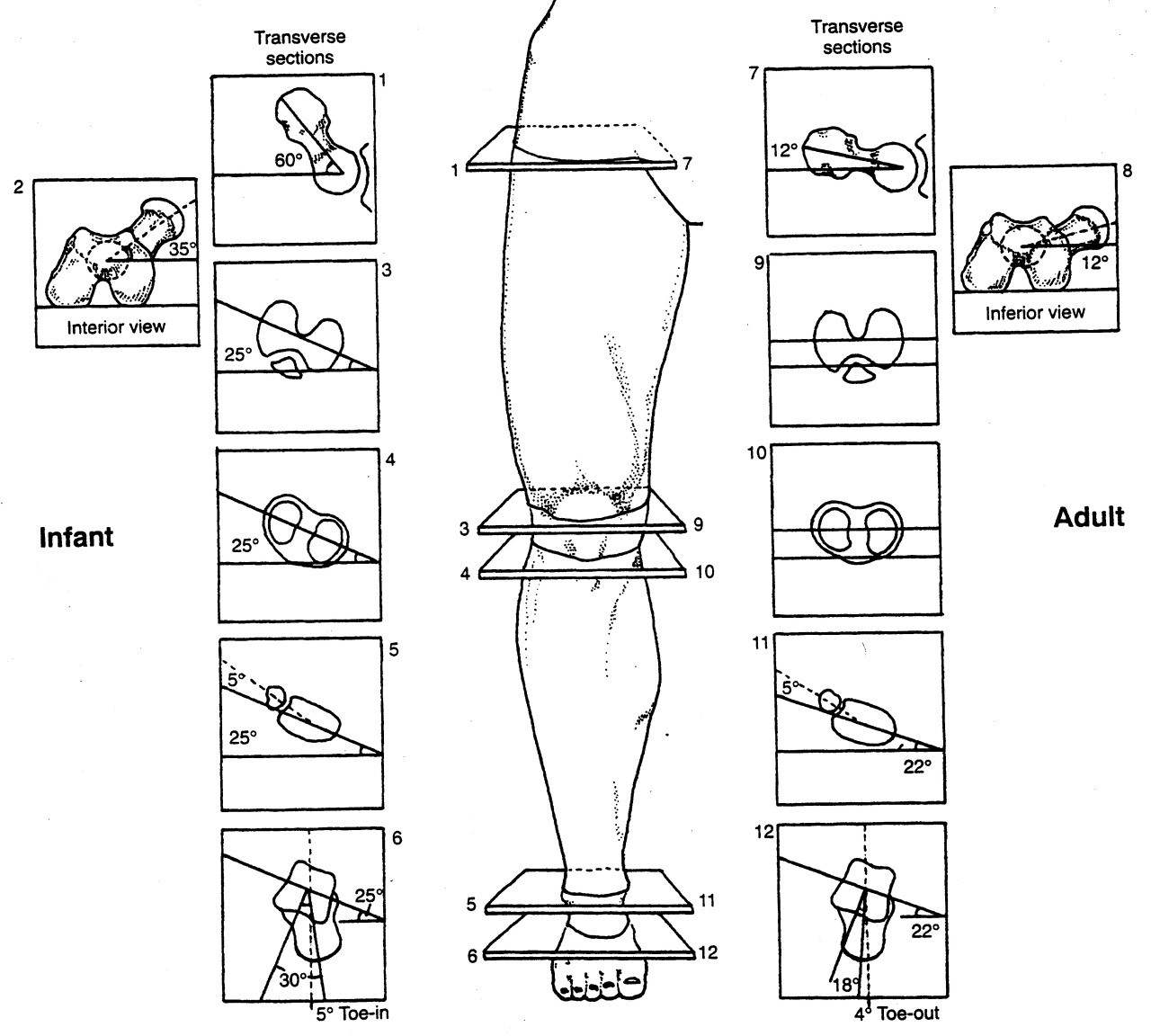
Looking at the top left picture: we can see that the axis of the tibial plateau and the transmalleolar axis (an imaginary line drawn through the medial and lateral malleolus) are parallel at birth (net angle zero) and progress to 22 degrees at skeletal maturity, resulting from the outward rotation of the tibia of about 1-1.5 degrees per year. This results in a normal external tibial version of about 17-18 degrees (you subtract 5 degrees for the talar neck angle, talked about in the link above). Note that this is the normal or ideal angle we would expect (hope?) to see. Go 2 standard deviations in either direction and we have external and internal tibial torsions.

You can go about taking this measurement in may ways; we will outline 2 of them. 

  1. In the upper left picture, we see an individual who has their knee flexed to 90 degrees over the side of a table while seated. This represents the tibial plateau angle. You the use a protractor to measure the angle between the tibial plateau and an imaginary line drawn through the medial and lateral malleoli. This is the transmalleolar angle. You then subtract 5 degrees from this number (remember the talar neck angle?) to get the angle of tibial version (or torsion).
  2. In the lower left and right pictures, we have the patient supine with the knees pointed upward and tibial plateau flat on the table. Then, working from inferiorly, use a goniometer to measure the angle of the transmalleolar axis. Again, we subtract 5 degrees for the talar neck.

We would encourage you to read up on torsions. This post, which we wrote over a year ago, is probably one of the most important ones on tibial torsions. 

Torsions. Important stuff, especially when you are talking about the axis of the knees in activities like a squat. Remember, the knee is a hinge between 2 multiaxial joints (hip and ankle) and will often take the brunt of the (patho)mechanics, as it has fewer degrees of freedom of movement. If you have external tibial torsion and you push your knees (angle your feet) out further, you are moving the knees outside the saggital plane. You have better have a very competent medial tripod! If you have internal tibial torsion, angling the feet out may be a good idea. Know your (or your patients/clients/athletes) anatomy!

The Gait Guys. Bald, Good Looking and Twisted. Here to help you navigate your way through better biomechanics. 

Podcast 72: Neuroplasticity, EVA Shoe Foam, and Shoe Trends

Maximalist shoes and the death of Minimalism ? Could this be true ?

*Show sponsor: www.newbalancechicago.com

Lems Shoes.  www.lemsshoes.comMention GAIT15 at check out for a 15% discount through August 31st, 2014.

A. Link to our server: 

http://traffic.libsyn.com/thegaitguys/pod_73f.mp3

Direct Download: 

http://thegaitguys.libsyn.com/podcast-72

B. iTunes link:

https://itunes.apple.com/us/podcast/the-gait-guys-podcast/id559864138

C. Gait Guys online /download store (National Shoe Fit Certification and more !) :

http://store.payloadz.com/results/results.aspx?m=80204

D. other web based Gait Guys lectures:

www.onlinece.com   type in Dr. Waerlop or Dr. Allen,  ”Biomechanics”

______________

Today’s Show notes:

1. Neuroplasticity: Your Brain’s Amazing Ability to Form New Habits
new link (does not have the old photo ivo mentioned that he loved)
 
2. Last week we pounded the sand on EVA foam and maximalist shoes. There was alot of attention, emails and good social media discussion on the topic.  
LETS REVIEW IT
file:///Users/admin/Downloads/p142_Heel_shoe_interactions_and_EVA_foam_f_web_150dpi.pdf
 
3. Then there just last week there was an article in LER on “the death of minimalist shoes” ? 
READ THIS: 
The rise and fall of minimalist footwear | Lower Extremity Review Magazine
http://lermagazine.com/cover_story/the-rise-and-fall-of-minimalist-footwear
 

4.  Physical Therapy as Effective as Surgery for Meniscal Tear

Kathleen Louden

March 20, 2013
Torn Meniscus? Thinking about surgery? Think again…

5. Cast study: the broken foot tripod
Invalid video embed.
Proprioceptive afferent inputs can control the timing and pattern of locomotion. When disease is present, or when injury has compromised the neuro-biomechanical linkages, slow postural responses can trump what timely responses are necessary to ensure for smooth locomotion.
 
When many people think of balance and locomotion, the cerebellum is often a top topic for it is important for movement control and plays a particularly crucial role. Thus, a most characteristic sign of cerebellar damage is walking ataxia. It is not known how the cerebellum normally contributes to walking, although recent work suggests that it plays a role in the generation of appropriate patterns of limb movements, dynamic regulation of balance, and adaptation of posture and locomotion through practice. (1)
Reflex pathways exist which regulate the timing of the transition from stance to swing, and control the magnitude of ongoing motoneuronal activity. During locomotion there is a closely regulated feedback from the various sensory receptors in the skin, joints, muscles, tendons, ligaments and other tissues, this is referred to as afferent feedback. When there is damage to these sensory “organs”, or the pathways into, or out of, the central nervous system locomotion becomes difficult.  We can see this in the video case above. This is a case of Chronic Inflammatory Demyelinating Polyradiculopathy (CIDP). It is an immunne-mediated inflammatory disorder of the peripheral nervous system whereby the myelin sheath of neurons is slowly eroded and as a result, the affected nerves and pathways fail to respond well rendering numbness, paresthesias, pain and progressive muscle weakness along with loss of deep tendon refexes. Obviously this will render locomotion fatiguing and difficult. Falls are not uncommon as you can see in the video.
 
Timing and coordination is everything in gait. When a portion of the system is compromised from injury or neurologic deficit, locomotion becomes strained.  There is an intricate balance between the extensor and flexor muscles.  We found this quote by Lam and Pearson particularly relevant to today’s discussion and video.

“Proprioceptive feedback from extensor muscles during the stance phase ensures that the leg does not go into swing when loaded and that the magnitude of extensor activity is adequate for support. Proprioceptive feedback from flexor muscles towards the end of the stance phase facilitates the initiation of the swing phase of walking. Evidence that muscle afferent feedback also contributes to the magnitude and duration of flexor activity during the swing phase has been demonstrated recently. The regulation of the magnitude and duration of extensor and flexor activity during locomotion is mediated by monosynaptic, disynaptic, and polysynaptic muscle afferent pathways in the spinal cord. In addition to allowing for rapid adaptation in motor output during walking, afferent feedback from muscle proprioceptors is also involved in longer-term adaptations in response to changes in the biomechanical or neuromuscular properties of the walking system.” (2)

Gait and any form of locomotion are highly complicated with many pieces necessary to achieve clean, smooth, coordinated motion.  Failure in only one piece of the puzzle can result in profound unhinging of the entire system because of the entangled nature of the feedback loops.  
Nothing dramatic today gang, just some thoughts that came to us after seeing this client and doing some reading to keep up on things.  We thought this would be a nice follow up to Monday’ blog post on proprioception.
Shawn and Ivo
the gait guys
References:
1. Neuroscientist. 2004 Jun;10(3):247-59.

Cerebellar control of balance and locomotion.

2. Adv Exp Med Biol. 2002;508:343-55.

The role of proprioceptive feedback in the regulation and adaptation of locomotor activity. Lam T1, Pearson KG.

tumblr_nahfwdDSeS1qhko2so1_1280.jpg
tumblr_nahfwdDSeS1qhko2so2_1280.jpg

Proprioception trumps Biomechanics

As I sit here on a rare Friday afternoon, not working (OK, I am writing this, so sort of working) and looking out at the lake (picture above), while on a family camping trip, I think about a walk on the rocks this morning with my kids. I was watching my very skilled 7 year old jump from rock to rock while my 3 ½ year old, that thinks he is seven, tried to follow his older brother.

I had my foot on a rock which lowered the front of my foot in plantar flexion and stood on that leg. I noticed that my balance was not as great as it was when my foot was in dorsiflexion. This made me think about pronation and supination. Yes, it is not uncommon for me to think about such things, especially when I have some spare time. That is one of the things about being a foot and gait nerd; these sorts of things are always on our minds.

So, why was my balance off? Did I need more proprioceptive work? Were my foot intrinsics having issues? No, it was something much more mundane.

Pronation consists of dorsiflexion, eversion and abduction. This places the foot in a  “mobile adaptor” posture, reminiscent of our hunter/gatherer ancestors, who needed to adapt to uneven surfaces while walking over terra firma barefoot. Supination, on the other hand (which is the position my foot was in), consists of plantarflexion, inversion and adduction. It places the foot (particularly the midtarsals) in a locked position for propulsion (think of the foot position during toe off).

So why when my foot was plantar flexed and adducted while standing on this rock so much more unstable in this supposedly more stable, supinated position? I would encourage you, at this point, to try this so you can see what I mean. When I placed my foot in dorsiflexion on the rock, I was much more stable. A most interesting conundrum for a biomechanist.

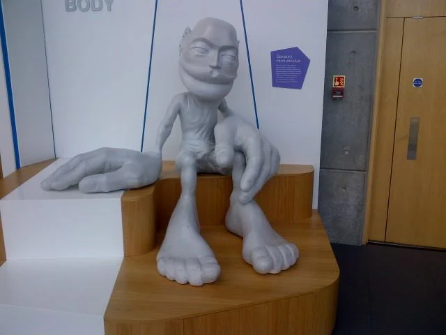
Experimenting for a few minutes, alternating plantar flexion and dorsi flexion, gave me the answer. When we are walking on the flats, our foot is (usually) not pushed to the extremes of dorsiflexion; with the front of the foot up on a rock, it is much more so. This “extra” upward force on the front of the foot, provides much more sensory input (and thus proprioception) from the ball of the feet. Take a look at the sensory homunculus and you can see how much brain real estate is dedicated to your foot, especially the front portion. With this information, we are able to apply more  force through the posterior compartment of the leg,which is stronger than my anterior compartment (as it is with most folks).

When the front of your foot is in plantar flexion (ie, your heel is on the rock), we have less sensory input to the balls of the feet, and rely more on the anterior compartment (weaker in many folks, including myself) to counterbalance the weight of our body.

Mystery solved: proprioception trumps biomechanics; more proof that the brain is smarter than we are.

The Gait Guys. Solving the worlds great gait questions, one at a time.

tumblr_n9t3r07m8O1qhko2so5_r1_1280.png
tumblr_n9t3r07m8O1qhko2so1_500.png
tumblr_n9t3r07m8O1qhko2so2_1280.jpg
tumblr_n9t3r07m8O1qhko2so3_250.jpg
tumblr_n9t3r07m8O1qhko2so4_250.jpg

Did you see this in our recent blog post here ? a reader made us look closer. Did you catch it ?
The clients right foot appears to have a dropped 1st met head. (we hate this term, because it is not accurate and is a sloppy clinical description). In this still photo it appears plantarflexed.  But in this video, consider the descended 1st met head as due to the disuse or weakness of the EHL muscle (extensor hallucis longus) of the 1st toe. Or, is this in fact a compensated forefoot varus ? Sure looks like it. But with all that anterior compartment weakness (as we discussed in the previous blog post link above) it could just be a mirage. In the photo above, in a normal foot the rearfoot plane (greenline) should parallel the forefoot line (orange line). In this case, in this actively postured foot (thus some inaccuracy here, we are merely making a teaching point from the photo) the upslope of the orange line suggests a forefoot varus. This would be true if the first Metatarsal head also was on this line, but you can see that it has its own idea. This represents, in theory (regarding this photo), a compensated forefoot varus. But remember, this client is  holding the foot actively in this posture. A true hands on assessment is needed to truly define a Forefoot varus, and whether it is anatomic, flexible, rigid or in many cases, just a learned functional posturing from weakness of the flexor/extensor pairing of the 1st metatarsal complex or from other weaknesses of the other forefoot evertors.  It gets complicated as you can see.

As always, knowledge of the anatomy and functional anatomy allows for observation, and observation leads to understanding, which leads to answers and then remedy implementation. Our thoughts, knowing the case, is that this is a functional appearance illusion of a compensated forefoot varus due to the EHL, EDL and tibialis anterior weakness (anterior compartment) and how they play together with the flexors. One must be sure to assess the EHL when examining the foot. Test all of the muscles one by one.  We have been talking about toe extensors for a long time, they can be a paramount steering wheel for the forefoot and arch posture. Podcast 71 talks about this Forefoot varus, and you should care.
In a 2009 study by Reynard et al they concluded: 

  • “The activity of extensor digitorum longus muscle during the swing phase of gait is important to balance the foot in the frontal plane. The activation of that muscle should be included in rehabilitation programs.” (1)

here is the video again.

Have a burning desire to learn more about forefoot varus, here are 25 blog post links from our last few years. And/or you can take our National Shoe Fit program (downloadable links below).

Knowing what you are seeing during your exam and gait analysis can only truly come from coupling your observations with a clinical exam.  Anything less is speculation and guess work.  It is gambling, and this is not Vegas baby, this is someone’s health.

Shawn and Ivo, The Gait Guys

________________

National Shoe Fit Certification Program:

Gait Guys online /download store (National Shoe Fit Certification and more !) :

http://store.payloadz.com/results/results.aspx?m=80204

1. Foot (Edinb). 2009 Jun;19(2):69-74. Epub 2008 Dec 31. Foot varus in stroke patients: muscular activity of extensor digitorum longus during the swing phase of gait.  Reynard F, Dériaz O, Bergeau J.

Other web based Gait Guys lectures:

www.onlinece.com   type in Dr. Waerlop or Dr. Allen,  ”Biomechanics”Reference

Podcast 71: Forefoot Varus, Big Toe Problems & Charlie Horses"

*Show sponsor: www.newbalancechicago.com

Lems Shoes.  www.lemsshoes.comMention GAIT15 at check out for a 15% discount through August 31st, 2014.

A. Link to our server: 

Direct Download: 

http://traffic.libsyn.com/thegaitguys/pod_72final.mp3

Permalink: http://thegaitguys.libsyn.com/podcast-71

B. iTunes link:

https://itunes.apple.com/us/podcast/the-gait-guys-podcast/id559864138

C. Gait Guys online /download store (National Shoe Fit Certification and more !) :

http://store.payloadz.com/results/results.aspx?m=80204

D. other web based Gait Guys lectures:

www.onlinece.com   type in Dr. Waerlop or Dr. Allen,  ”Biomechanics”

______________

Today’s Show notes:

1. American College of Cardiology. Running out your healthy heart. How much exercise is too much ?

Running for 7 minutes a day cuts risk of death by 30%, study says
http://wgntv.com/2014/07/29/running-for-7-minutes-a-day-cuts-risk-of-death-by-30-study-says/
 
2. The history of “Charlie Horses”
 
3. A runner with strange shin bruises.  
from : Joy 
Hi, I’m a great follower of your blog - fascinating stuff! I was wondering if I could ask you a quick question as nobody I’ve spoken to has been able to help:

I’ve been getting bruises that appear on my shin during running. They don’t hurt, I’m just wary of ignoring what could be a warning sign. Have you ever come across this before? (It’s mainly the spot where I had a tibial stress fracture last year, but I also get a few other apparently spontaneous bruises on my lower legs.)
4. Is that a forefoot varus or are you just happy to see me ?
Functional vs Anatomic vs. Compensated forefoot varus foot postures. A loose discussion.
5. A reader’s pet peeve about shoe store “gait analysis”.
6. Thoughts on pronation and the like.
7. Case study:  First toe fusion and implications long and short term.
“I had a patient today with an MTP fusion of his great toe after adverse complications from a bunionectomy.  Do you have any recommendations for gait training when great toe dorsiflexion is no longer an option?  He is currently compensating by externally rotating his foot and overpronating.  I’m thinking through it and  I know he has to gain the motion elsewhere to help normalize his gait as much as possible, so possibly gaining ankle dorsiflexion and hip extension.  Just wondering if you have any tips to share or articles to point me to for further ideas.  Continuing my research now.  I’m a relatively new grad and this is my first patient I’m seeing with this fusion. Many thanks
tumblr_n9jt5dtWlc1qhko2so1_1280.jpg
tumblr_n9jt5dtWlc1qhko2so2_1280.jpg
tumblr_n9jt5dtWlc1qhko2so3_1280.jpg
tumblr_n9jt5dtWlc1qhko2so4_r1_1280.gif

Subtle clues. Helping someone around their anatomy

This patient comes in with low back pain of years duration, helped temporarily with manipulation and activity. Her exam is relatively benign, save for increased lumbar discomfort with axial compression in extension and extension combined with lateral bending. Believe it or not, her abdominal and gluteal muscles (yes, all of them) test strong (no, we couldn’t believe it either; she is extremely regular with her exercises). She has bilateral internal tibial torsion (ITT) and bilateral femoral retro torsion (FRT). She has a decreased progression angle of the feet during walking and the knees do not progress past midlilne. There is a loss of active ankle rocker with gait, but not on the exam table; same with hip extension. 

We know she has a sweater on which obscures things a bit, but this is what you have to work with. Look carefully at her posture from the side. The gravitational line should pass from the earlobe, through the shoulder, greater trochanter and through or just anterior to the lateral malleolus.

In the top picture, can you see how her pelvis is anterior to this line? Do you see how it gets worse when she lifts her hands over her head (yes, they are directly over head)? This can signify many things, but often indicates a lack of flexibility in the lumbar lordosis; in this case, she cannot extend her lumbar spine further so she translates her pelvis forward. Most folks should have enough range of motion from a neutral pelvis and enough stability to allow the movement to occur without a significant change. Go ahead, we know you are curious, go watch yourself do this in a mirror and see if YOU change.

Looking at the bottom left picture, can you pick out that she has a genu valgus? Look at the hips and look at the tibial angle.

In the bottom left picture, did you note the progression angle (or lack of) in her feet? This is a common finding (but NOT pathognomonic) in patients with internal tibial torsion. Notice the forefoot adductus on the right foot?

So what do we think is going on?

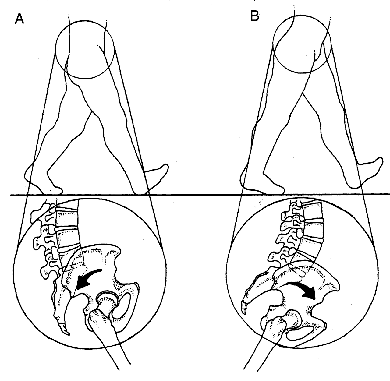
  • ITT and FRT both limit the amount of internal rotation of the thigh and lower leg. Remember you NEED 4 degrees of each to walk normally. Most folks have significantly more
  • if you don’t have enough internal rotation of the lower extremity, you will need to “create” it. You can do this by extending the lumbar spine (bottom picture, right) or externally rotating the lower extremity
  • Since her ITT and FRT are bilateral, she flexes the pelvis and nutates the pelvis anteriorly.
  • the lumbar facet joints should only carry 20% of load
  • she is increasing the load and causing facet imbercation resulting in LBP.

What did we do?

  • taught her about neutral pelvic positioning, creating more ROM in the lumbar spine
  • had her consciously alter her progression angle of her foot on strike, to create more available ROM in internal rotation
  • encouraged her to wear neutral shoes
  • worked on helping her to create more ankle rocker and hip extension with active drills and exercise (ie gait rehabilitation); shuffle walks, Texas walk, toes up walking, etc

why didn’t we put her in an orthotic to externally rotate her lower extremity? Because with internal tibial torsion, this would move her knee outside the saggital plane and create a biomechanical conflict at the knee and possibly compromising her meniscus.

Cool case, eh? We thought so. Keep on learning so your brain keeps expanding. If you are not growing your brain, you are shrinking it!

The Gait Guys

Here is a great case from a reader.


“Hey guys, I absolutely love the show, especially as it becomes less and less over my head. Due to your love of gait-altering absurdly thick EVA midsoles, I thought you might like to check out this Hoka in…

Here is a great case from a reader.

“Hey guys, I absolutely love the show, especially as it becomes less and less over my head.

Due to your love of gait-altering absurdly thick EVA midsoles, I thought you might like to check out this Hoka incident that occurred at the Marathon des Sable across the Sahara in Morocco, a 6 day 251km event. It was posted by Ian Corless at Talk Ultra Podcast. Apparently the medial side of the midsole collapsed–on DAY 2! This guy finished the race, and as you have to carry 100% of your gear and nutrition, I guess he only had the one pair. It looks like this runner should fly out to CO or IL asap, because if he didn’t have gait issues before, he is sure to have them now.”

This brings up some scary thoughts when it comes to the amount of EVA foam and quality of foam (EVA or otherwise) being used in some shoes.  "The more foam there exists, the greater one can break down into their compensation or deforming strategy.“ What do we mean by this ?  Well, two things should be on one’s mind:  1. all foam breaks down into the vector of the deforming forces and 2. most of us do not have perfectly clean biomechanics, thus an abnormal loading vector is most likely present. These aberrant biomechanics are eventually reflected into our shoes as a "wear pattern”.  In this case, the EVA foam had progressively broken down into their rearfoot pronation (and likely mid and forefoot pronation). In this case, even if the person had enough tibialis posterior and other medial pronation-decelerating structure strength at the start, the acceleration of their foot into this issue is now even more abrupt, brisk, excessive etc.  A new pair of shoes would not be broken down into this deformity and so a newer pair of shoes is preventive. This is why we recommend new shoes often, and the cycling in of another pair (or several pair) into the mix so that one is never driving the same shoes into the potentially destructive compensation patterns that most of us  have.  At least with a fresh pair of shoes brought into the mix at the 200 mile wear point, you would only be in the more destructive shoes every other run, giving the body time to recuperate more. 

As for this pair of shoes, this runner either has a terrible right foot problem or this was a brutally flawed right shoe from the get go, or both. We can only imaging how painful the medial knee might be at this point.  Furthermore, imagine the abrupt nature of the hip internal rotation mechanics ! IF they do not have hip labrum impingement yet, they will soon !  And with that amount of internal limb spin, can you imagine how inhibited the glutes would be from constantly having to eccentrically control that excessive rotation? 

As a whole, are not huge fans of the HOKA shoe family, we just cannot fathom the need for this much foam under the feet. If you have been with us long enough you will have heard on our podcast and blog talk about increased impact forces with increasing EVA foam thickness (want that info, here is the link and references). Just because some EVA foam is good, doesn’t mean more is better.  Remember, to propulse off of a foam infrastructure you must bottom out/compress the foam sufficiently to find a firmness to propulse from. The Hoka’s have plenty of foam making this our concern, and we are not picking on just them. There are other companies doing this “super sizing/super stacking” such as Brooks, Altra, and New Balance to name just a few.  Sure they have added a greater forefoot rocker/toe spring on the front of the shoe to help (they have to because the foam thickness is so great that there is no flexing of the forefoot of the shoes), but is it enough for you? Remember, every biomechanical phase of the gait cycle is necessary and timely to engage the natural joint, ligament, muscle components of joint loading, mobility, stability and movement. If you spend too much time in one phase (perhaps because you are waiting for foam to decompress) you may wait a moment too long and miss the opportunity for another critical phase to begin in the sequence.  This is the root cause of many injuries, aberrant biomechanics leading to aberrant mobility or stability. 

So remember these few things:

1. more is not always better for you, it may be for some, but maybe not you.

2. there is a price to pay somewhere in the mechanical system, after all the body is a contained system. What doesn’t happen at one joint often has to be made up at the next proximal or distal joint.

3. Everyone has some aberrant mechanics. No one is perfect. These imperfections will reflect in your shoes, and the longer you are in a pair of shoes the deeper the aberrant mechanics will be reflected in your shoe, thus acting as a steering wheel for the aberrant pattern (the steering is more direct/ more aggressive than in a new pair of shoes). So keep at least 2 pair of shoes rotating in your run cycle, one newer and one half done. We even recommend 3 pairs often.  Trust us, the sudden biomechanical shift from a dead shoe into a new one (even though it is a clean new shoe without bad patterns in it) is still a biomechanical shift and could cause adaptive phase problems, pain or injury.

Lots to consider in this game. It is not just about dropping into barefoot and taking off down your street. Not if you want to be doing this for a long time and stay healthy.

Shawn and Ivo, the gait guys

* next day follow up from our social media pages:

Along the lines of EVA and yesterdays post: 

“Wear of the EVA consistently increased heel pad stresses, and reduced EVA thickness was the most influential factor, e.g., for a 50% reduction in thickness, peak heel pad stress increased by 19%. ”

This study looks at a model; it would be interesting to see this study with a large cohort.

Biomed Mater Eng. 2006;16(5):289-99.

Role of EVA viscoelastic properties in the protective performance of a sport shoe: computational studies.

Even-Tzur N1, Weisz E, Hirsch-Falk Y, Gefen A.

http://www.ncbi.nlm.nih.gov/pubmed/17075164

tumblr_n9ql3rlqGi1qhko2so2_r1_1280.jpg
tumblr_n9ql3rlqGi1qhko2so1_1280.gif
tumblr_n9ql3rlqGi1qhko2so3_r1_1280.gif
tumblr_n9ql3rlqGi1qhko2so4_r1_1280.gif

ETT and Hip Extension

Not Extra Terrestrial Tricks, but rather External Tibial Torsion. How it effects hip extension.

We received this question from Matthew P on our Facebook post from 8/1 (original post from here) which was based on this article, and thought it would make an excellent opportunity to teach. 

I looked at this yesterday and had actually first come across it a year or more ago when I was trying to find some resources for femoral torsion. You guys are about the only ones discussing at length the impact and implication of adult femoral torsion.

Re: tibial torsion and your post yesterday saying that > 30deg external torsion can affect both knee and hip extension, what I still don’t understand is the mechanism behind the hip limitation. How would that torsion translate through the leg to the hip?

There are a few things we need to remember to make sense of this:

  • tibial torsion is the angular difference between the tibial plateau and distal tibial malleoli and refers only to the tibia, not the entire lower extremity (see top photo)
  • pronation can occur in the rear foot, mid foot, and fore foot
  • pronation causes internal spin of the leg and thigh, due to plantar flexion, eversion and abduction of the talus (see middle photo)
  • internal spin of the hip causes posterior translation of the femoral head via the “glide and roll” phenomenon
  • these are appropriate mechanics during the 1st ½ of the gait cycle (initial contact to mid stance)
  • pronation is one of the 4 shock absorbing mechanisms (pronation, ankle dorsiflexion, knee flexion, hip flexion) of the lower extremity

now try this (yes, at home!)

  • flex your knee
  • internally rotate you leg
  • allow your arch to flatten
  • try and extend your hip

Remember these facts about supination

  • supination is initiated by the swing phase leg as it starts in early swing and continues to terminal swing (see third picture)
  • supination (from full pronation), should occur from midstance to pre swing
  • supination makes the foot into a “rigid lever” to transpose forces from above the foot into the foot and allow for propulsion (see third picture)
  • supination involves external rotation of the lower leg and thigh (see pictures 3 and 4)
  • external rotation of the hip is accompanied by anterior glide of the femoral head via the “glide and roll” phenomenon
  • this position puts the gluteal muscles (max and posterior fibers of the gluteus medius) at a mechanical advantage

now try this:

  • extend your knee
  • externally rotate your leg
  • you should have a full arch at this point
  • try and extend your hip

What did you (hopefully) learn?

  1. When the hip is in an externally rotated position it is easier to extend; the femur head moves anteriorly, the femoral joint capsule becomes tighter and stability is created
  2. when the hip is internally rotated, it is more difficult to extend
  • the femur head glides posteriorly, changing the axis of rotation of the joint
  • the gluteus maximus and posterior fibers of the gluteus medius are at a mechanical disadvantage

OK. Got it? We sure hope so! Excellent question, Matthew. Thanks for the opportunity to teach this concept.

The Gait Guys. Taking you closer to mastering the gait cycle with each post. 

Podcast 70: Achilles Solutions and Foot Cases

The Achilles and Calf: Achieve Posterior Length via Anterior Strength

A. Link to our server: 

Direct Download: 

http://traffic.libsyn.com/thegaitguys/pod_70final_was71.mp3

Permalink: http://thegaitguys.libsyn.com/podcast-70-0B. 

iTunes link:

https://itunes.apple.com/us/podcast/the-gait-guys-podcast/id559864138

C. Gait Guys online /download store (National Shoe Fit Certification and more !) :

http://store.payloadz.com/results/results.aspx?m=80204

D. other web based Gait Guys lectures:

www.onlinece.com   type in Dr. Waerlop or Dr. Allen,  ”Biomechanics”

______________

Today’s Show notes:

*Show sponsor: Lems Shoes.  www.lemsshoes.com
Mention GAIT15 at check out for a 15% discount through August 31st.
 
1. Achilles tendons, loading, and biomechanical changes with different shoes and heel stack heights.
 
2. Aging adults, falls and keeping them and their gait safe.
 
3. Gait and speed evolution of vertebrates.
 
4. Blaise Dubois et al on Barefoot Running. Shod vs unshod.
 
5. Females, pronation, and back pain. The Framingham foot study.
 
6. Your feet and orienteering.
 
7. A case of calcaneal valgus in a youngster.
 
8. Structural integrity is decreased in both Achilles tendons in people with unilateral Achilles tendinopathy
http://www.jsams.org/article/S1440-2440(14)00115-7/abstract