Podcast 90: The brain: A deeper look at gait and motor control

Show sponsors:
www.newbalancechicago.com


Other Gait Guys stuff

A. Server links to the podcast:

http://directory.libsyn.com/episode/index/id/3302518

http://traffic.libsyn.com/thegaitguys/pod_90f.mp3

http://thegaitguys.libsyn.com/90

B. iTunes link:
https://itunes.apple.com/us/podcast/the-gait-guys-podcast/id559864138

C. Gait Guys online /download store (National Shoe Fit Certification and more !) :
http://store.payloadz.com/results/results.aspx?m=80204

D. other web based Gait Guys lectures:
Monthly lectures at : www.onlinece.com   type in Dr. Waerlop or Dr. Allen,  ”Biomechanics”

Our Book: Pedographs and Gait Analysis and Clinical Case Studies

electronic copies available here:

Amazon/Kindle:
http://www.amazon.com/Pedographs-Gait-Analysis-Clinical-Studies-ebook/dp/B00AC18M3E

Barnes and Noble / Nook Reader:
http://www.barnesandnoble.com/w/pedographs-and-gait-analysis-ivo-waerlop-and-shawn-allen/1112754833?ean=9781466953895

https://itunes.apple.com/us/book/pedographs-and-gait-analysis/id554516085?mt=11

Hardcopy available from our publisher:
http://bookstore.trafford.com/Products/SKU-000155825/Pedographs-and-Gait-Analysis.aspx


Show notes:

Researchers Identify Important Control Mechanisms for Walking
http://neurosciencenews.com/spinal-cord-activation-neurology-walking-1698/
 “Using statistical methods, we were able to identify a small number of basic patterns that underlie muscle activities in the legs and control periodic activation or deactivation of muscles to produce cyclical movements, such as those associated with walking

People watching: Different brain pathways responsible for person, movement recognition
http://medicalxpress.com/news/2015-01-people-brain-pathways-responsible-person.html
Each time you see a person that you know, your brain seemingly effortlessly and immediately recognizes that person by his or her face and body. Just as easily, your brain understands a person’s movements, allowing you to perform critical skills such as interpreting social cues, detecting threats and determining the difference between skipping and jumping.

Remapping the brian.
http://news360.com/article/273798702/#
Published in the Journal of Neuroscience, 

How Rotation gets you dorsiflexion: Easy solutions for ankle mobility
 http://thenominalist.wordpress.com/2014/12/16/how-rotation-gets-you-dorsiflexion-easy-solutions-for-ankle-mobility/

Wedges: http://www.footfoundation.com/

Its hard to change the neurology of engrained habits……..
Neuromuscular Exercise Post Partial Medial Meniscectomy: Randomized Controlled Trial.
Hall M1, Hinman RS, Wrigley TV, Roos EM, Hodges PW, Staples MP, Bennell KL.
Journal Med Sci Sports Exerc. 2014 Dec 23. [Epub ahead of print]

Doc Martins Boots: Tobias and Curtis
http://www.journeys.com/product.aspx?id=243165&green=157F2C73-F732-5ACF-AC09-89894E6EE1F1
Tobias and Curtis

Relationship between physical impairments and movement patterns during gait in patients with end-stage hip osteoarthritis
Joseph Zeni, Jr.1,2  et al
http://onlinelibrary.wiley.com/doi/10.1002/jor.22772/abstract;jsessionid=E0C246BA281C71A4124C03FEB608C474.f02t02

VAncouver Gait course: http://twinbridgesphysiotherapy.com/courses-events/foot-and-gait-course/

Prof. Ted Carrick and the eyes, and some cursory thoughts on gait and brain function as a whole.  We have been blessed to learn from this man and those from his institute, come listen and find out why.

The movements of the eyes are keys to human brain function and movement such as gait.  What kind of eye stuff you ask ? 

How are your clients eye pursuits, saccades, VOR, vergence, OPK or fixation abilities ? All 6 of these are necessary for normal eye and brain function. Without these working properly gait can also be impaired and muscles will not function correctly if they are tied directly to the gait and movement systems.

This is just the tip of the ice berg however.  What about the function of your client’s basal ganglia, thalamus, cerebellum, mesencephalon, cortex, or the vestibular system, as a small sampling. What about the tracts that feed and interconnect all of this stuff, like the corticospinal, vestibulocerebellar, spinocerebellar, rubrospinal, recticulospinal, or vestibulospinal tracts, to name a few ? What about the lobes of the brain, the frontal, parietal, occipital, temporal ?  

Dear gait brethren, you must see that human function is about the nervous system. Nothing happens to the end organ receptors, the muscles, joints, motor patterns and others without proper orchestration of the central, peripheral and autonomic systems. Gait is nothing short of a miraculous event bringing all of the nervous system’s amazing parts into a beautiful symphony of timed and rhythmic events, arm swing, balance, vision, proprioception, postural restrain from gravity and so much more. 

Don’t get too caught up in the latest greatest treatment fad or exercise on the web without understanding that safe, effective, efficient, pain free human locomotion is a product of the orchestra’s grand conductor, the brain.  
The brain is organized beautifully. Do you find yourself over and over again activating your client’s proximal flexors ? You are plugging into the rubrospinal pathways, and perhaps that is not where the golden honey and buscuits are found.  And if you find yourself delving into your client’s distal extensors ? Well, you are plugging into their recticulospinal pathways. How about their proximal extensors ? … . lateral vestibulospinal pathways.  Treatment cannot, and should not, be random. There is a recipe and a right way.  You are either part of your client’s solution or part of their problem. 

Thank you for your brilliance Dr. Ted Carrick, you have changed our lives and those that want the deeper answers as to why and how.  When you know these answers, you don’t need to dip into the latest greatest super double chocolate fudge brownie ice cream “exercise” of the week, when cool and calculated pure Vanilla bean at the right place and the right time will serve as the best answer … .  if you know what you are dealing with, and if you have the right tools.

More on this fun stuff another time. Have a great week gait brethren !

Shawn and Ivo

the gait guys

tumblr_nhdevsLYyN1qhko2so3_400.jpg
tumblr_nhdevsLYyN1qhko2so4_r1_250.jpg
tumblr_nhdevsLYyN1qhko2so1_1280.jpg
tumblr_nhdevsLYyN1qhko2so2_250.jpg

Keeping it Objective.

For clinicians and some die hard foot geeks, we often like to keep things objective. What could be more objective than an angular measurement? A few important measurements when examining or radiographing feet can give us information about clinical decision making (not that we suggest radiographs for mensuration purposes unless you are a surgeon, but when they are already available, why not put them to good use ?). When things fall outside the accepted range, or appear to be heading that way, these numbers can help guide us when to intervene. 

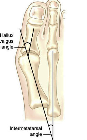
Hallux valgus refers to the big toe headed west (or east, depending on the foot and your GPS). In other words, the proximal and distal phalanyx of the great toe (hallux) have an angle with the 1st metatarsal shaft of typically > 15 degrees. This angle, called the Hallux Valgus Angle (HVA above) is used to judge severity, often for surgical intervention purposes but can guide conservative management as well. 

Metatarsus Primus Varus (literally, varus deformity of the 1st metatarsal) often accompanies Hallux Valgus. It describes medial deviation of the 1st metatarsal shaft, greater than 9 degrees. This angle is called the intermetatarsal angle and is measured by the angle formed by lines drawn parallel along the long axis of the 1st and 2nd metatarsal shafts. 

One other measurement is the Distal Metatarsal Articular Angle, which measures the angle between the metatarsal shaft and the base of the distal articular cap (ie, where the cartilage is) of the 1st metatarsal. This typically should be less than 10 degrees, preferably less than 6 degrees. Remember, these are static angles, things can change with movement, engagement, weight bearing strategies and shoes. What you see statically does not always predict dynamic angles and joint relationship.s

Are you doing surgery? Perhaps, as a last resort. Hallux valgus and metatarsus primus varus can be treated conservatively.

How do you do that?

The answer is both simple and complex.

The simple answer is: anchor the head of the 1st ray and normalize foot function. This could be accomplished by:

  • EHB exercises to descend the head of the 1st metatarsal
  • exercise the peroneus longus, to assist in descending the head of the 1st metatarsal
  • short flexor exercises, such as toe waving, to raise the heads of the lesser metatarsals relative to the 1st
  • work the long extensors, particularly of the lesser metatarsals to create balance between the flexors and extensors
  • consider using a product like “Correct Toes” to normalize the pull of the muscles and physically move the proximal and distal phalanyx of the hallux
  • wear shoes with wide toe boxes, to allow the foot to physically splay
  • consider using an orthotic with a 1st ray cut out, to help descend the head of the 1st metatarsal

This is by no means an exhaustive list and you probably have some ideas of your own. 

The complex answer is that in the above example, we have only included conservative interventions for the foot and have not moved further up the kinetic (or neurological chain). Could improving ankle rocker help create more normal mechanics? Would you accomplish this by working the anterior leg muscles, the hip extensors, or both? Could a weak abdominal external oblique be contributing? How about a faulty activation pattern of the gluteus medius? Could a congenital defect or genetic be playing a role? We have not asked “What caused this to occur in the 1st place?”

Examine your patients and clients. Understand the biomechanics of what is happening. Design a rehab program based on your findings. Try new ideas and therapies. it is only through our failures that we can truly learn.

The Gait Guys

references used:

http://www.bjjprocs.boneandjoint.org.uk/content/90-B/SUPP_II/228.3

http://www.slideshare.net/ANALISIS/hallux-valgus-2008-pp-tshare

http://www.orthobullets.com/foot-and-ankle/7008/hallux-valgus

http://www.slideshare.net/bahetisidharth/hallux-valgus-31768699?related=1

To met pad or not to met pad; that is the question. 

We use these in practice all the time. Think of it as a “helper” while you are training your patient or client to have better foot mechanics. 

“It’s all about the stress transfer, so what you’re trying to do is unload a certain spot on the foot—generally the met head with a met pad—and transfer it to a more proximal region,” 

Here is a nice evidence based review:

http://lermagazine.com/cover_story/evidence-based-use-of-metatarsal-pads
Social media reader comments and questions:
Reader: You demonstrated a peroneal exercise a few days back. In addition to the use of MT pads, is it effective in supporting transverse metatarsal arch?
  • The Gait Guys It can be. In our opinion, the exercises are key and the pad supports the exercises
  • The Gait Guys the goal is to restore foot function so that you do not need the pad. If your client is patient and willing to work on the exercises you likely won’t need a met pad, they may help speed things up, but remember, it is a crutch and crutches can be helpful but one can get lazy in using them and not do the supportive work. We only try to use them sparingly and only when necessary.

Following up on the peroneal topic of the week. HEre is a trademark exercise of ours.
http://thegaitguys.tumblr.com/post/63377772517/trade-secret-proper-calf-raise-we-are-selling

  • From a Reader: Just used the exercise on myself and a patient. Really like it. 2 questions. Can you get a better effect if performed on the edge of a step? Secondly, do you want to start in a tripod position and perhaps with short foot?
  • The Gait Guys We are assuming you mean letting the heel drop lower than the step/forefoot: and based on that assumption……if you feel that your client is ready for eccentric strength you can opt to do a slow decent into a negative heel. However, if there is ankle rocker limitation it would be wise to keep in mind that some of that end range dorsiflexion one might think they are getting from the ankle mortise joint might be from arch/midfoot collapse dorsiflexion , rear foot eversion and strain the plantar structures. Perhaps as they progress and gain still and control perhaps it is a good option. AWESOME question. As always, just because someone can do something doesnt mean they can do it safely. As for the tripod question…….well, they will be in the forefoot bipod mostly……bc the heel is off the ground……but this brings up technical issues if they are forefoot valgus or FF varus…..esp if they are rigid deformities……..it is doubtful many people are even considering this……..so once again, bravo Sir ! Is is quite often the small things that are the defining issues of the success or failure of a methodology. Bravo. !!!!! So glad you are part of this brethren David !

The Fudge Factor

The Fudge Factor

It is often easier to accomplish a task faster, rather than slower (like an exercise or skiing) because of the cortex “interpolating” or making its “best guess” as to what (based on experience) is going to happen. There is a certain amount of guess work (or what we call “the fudge factor”) involved.

Walking at a slower speed (or performing an exercise at a slower speed for that matter) has increased muscular demands, than doing it more quickly. Here is one study that exemplifies that.

“These findings may reflect a relatively higher than expected demand for peroneus longus and tibialis posterior to assist with medio-lateral foot stability at very slow speeds”

Gait Posture. 2014 Apr;39(4):1080-5. doi: 10.1016/j.gaitpost.2014.01.018. Epub 2014 Feb 6.
Electromyographic patterns of tibialis posterior and related muscles when walking at different speeds.
Murley GS1, Menz HB2, Landorf KB2.

The effect of walking speed on superficial lower limb muscles, such as tibialis anterior and triceps surae, is well established. However, there are no published data available for tibialis posterior - a muscle that plays an important role in controlling foot motion. The purpose of this study was to characterise the electromyographic timing and amplitude of selected lower limb muscles across five walking speeds. Thirty young adults were instructed to walk barefoot while electromyographic activity was recorded from tibialis posterior and peroneus longus via intramuscular electrodes, and medial gastrocnemius and tibialis anterior via surface electrodes. At faster walking speeds, peak electromyographic amplitude increased systematically during the contact and midstance/propulsion phases. Changes in the time of peak amplitude were also observed for tibialis posterior, tibialis anterior and peroneus longus activity; however, these were muscle and phase specific. During contact phase, peak electromyographic amplitude for tibialis posterior and peroneus longus was similar across very slow to slow walking speeds. During midstance/propulsion phase, peak electromyographic amplitude for tibialis posterior and medial gastrocnemius was similar across very slow to slow walking speeds. These findings may reflect a relatively higher than expected demand for peroneus longus and tibialis posterior to assist with medio-lateral foot stability at very slow speeds. Similarly, peak amplitude of medial gastrocnemius was also relatively unchanged at the very slow speed, presumably to compensate for the reduced forward momentum. The data presented in this study may serve as a reference for comparing similarly matched participants with foot deformity and/or pathological gait.

Copyright © 2014 Elsevier B.V. All rights reserved.

‪#‎gait‬
‪#‎thegaitguys‬

So you think you are an iliotibial band syndrome guru ?  This study has some interesting provoking thoughts about the mechanics we have all previously assumed. It is good to challenge established teachings, for it is only through interrogating old w…
So you think you are an iliotibial band syndrome guru ?  This study has some interesting provoking thoughts about the mechanics we have all previously assumed. It is good to challenge established teachings, for it is only through interrogating old ways that we may see the true light of things.
The iliotibial band (ITB) syndrome is a common overuse injury that is commonly misunderstood. It has been regarded as a friction syndrome where the ITB rubs against he lateral femoral epicondyle because of its previously assumed variable function, below 30 degrees knee extension it has been though to act as an extensor of the knee, and above 30 degrees (ie more knee flexion) it has been thought to act as flexor.  It is thought to be a culprit (with the biceps femoris) of the shift phenomenon in the “pivot shift test” for posterolateral rotatory instability of the knee (PLRI).  Here is an interesting perspective from a 2006 journal article.
“In all cadavers, the ITB was anchored to the distal femur by fibrous strands, associated with a layer of richly innervated and vascularized fat. In no cadaver, volunteer or patient was a bursa seen. The MR scans showed that the ITB was compressed against the epicondyle at 30° of knee flexion as a consequence of tibial internal rotation, but moved laterally in extension. MR signal changes in the patients with ITB syndrome were present in the region occupied by fat, deep to the ITB. The ITB is prevented from rolling over the epicondyle by its femoral anchorage and because it is a part of the fascia lata. We suggest that it creates the illusion of movement, because of changing tension in its anterior and posterior fibres during knee flexion. Thus, on anatomical grounds, ITB overuse injuries may be more likely to be associated with fat compression beneath the tract, rather than with repetitive friction as the knee flexes and extends.”

We found this article interesting because it challenges many thoughts about its actual movement, (“it creates an illusion of movement”) because of changing of tension in the anterior and posterior fibres. As this article suggests, it is unlikely that there is any forward and backward motion of the band over the epicondyle during flexion and extension, rather the illusion of movement is from a gradual shifting of load to and from the anterior and posterior fiber bundles during flexion/extension. It is also an interesting article to us because it suggests and challenges that the clinical phenomenon is associated with fat compression rather than friction over the epicondyle.  The authors go into discussion of how the fat beneath the distal ITBand at the knee level is well vascularized and that Pacinian corpuscles can be present in adipose tissue supporting the view that fat compression may have a proprioceptive role and a roll in pain production when the corpuscles undergo hypertrophy in such a clinical setting.
Just remember what we have been saying all along when treating what you think are lateral chain problems, the ITBand receives most of the tendon of the gluteus maximus so do not forget to examine the hip and pelvis function, but so not forget the critical contribution that impaired foot and ankle function can have proximally at the knee.
This study has some interesting provoking thoughts about the mechanics we have all previously assumed. It is good to challenge established teachings, for it is only through interrogating old ways that we may see the true light of things.
If you are looking for more of our thoughts on this topic, we discussed a clinical case in our last podcast (link here). 
Shawn and Ivo, 
the gait guys

Fairclough J, Hayashi K, Toumi H, et al. The functional anatomy of the iliotibial band during flexion and extension of the knee: implications for understanding iliotibial band syndrome. Journal of Anatomy 2006;208(3):309-316. doi:10.1111/j.1469-7580.2006.00531.x

CAI: More on Chronic Ankle Instability.

More peroneii action! In folks with chronic ankle instability, it contracts earlier, longer (throughout stance phase) but not stronger…This article looks at activation times and patterns of folks with chronic ankle instability. 

One should never wonder why repeated ankle sprains occur. We have hit this topic hard in the past.  Chronic Ankle Instability (CAI) clients exhibit prioprioceptive and postural control challenges. According to this article, additionally, CAI clients have gait. 

Time of activation for all muscles tested occurred earlier in the CAI group than in the control group. The peroneus longus was activated for a longer duration across the entire stride cycle in the CAI group.”

Individuals with CAI demonstrated neuromuscular-activation strategies throughout the lower extremity that were different from those of healthy control participants. ”

Did you see our trademark “goto” exercise in yesterday’s social media Facebook blog post ?  It is a keeper if you ask us.  Don’t ignore chronic peroneal challenges, they will come back to haunt you.

_________

Lower Extremity Muscle Activation in Patients With or Without Chronic Ankle Instability.  Mark A. FegerMEd, ATCLuke DonovanMEd, ATCJoseph M. HartPhD, ATCJay HertelPhD, ATC, FNATA, FACSM Department of Kinesiology, The University of Virginia, Charlottesville

http://www.natajournals.com/doi/abs/10.4085/1062-6050-50.2.06 

Results:  Time of activation for all muscles tested occurred earlier in the CAI group than in the control group. The peroneus longus was activated for a longer duration across the entire stride cycle in the CAI group (36.0% ± 10.3%) than the control group (23.3% ± 22.2%; P = .05). No differences were noted between groups for measures of electromyographic amplitude at either preinitial or postinitial contact (P > .05).

Conclusions:  We identified differences between the CAI and control groups in the timing of muscle activation relative to heel strike in multiple lower extremity muscles and in the percentage of activation time across the entire stride cycle in the peroneus longus muscle. Individuals with CAI demonstrated neuromuscular-activation strategies throughout the lower extremity that were different from those of healthy control participants. Targeted therapeutic interventions for CAI may need to be focused on restoring normal neuromuscular function during gait.

More research on the peronei and chronic ankle instability.

More peroneii action! In folks with chronic ankle instability, it contracts earlier, longer (throughout stance phase) but not stronger…This article looks at activation times and patterns of folks with chronic ankle instability. 

One should never wonder why repeated ankle sprains occur. We have hit this topic hard in the past. Chronic Ankle Instability (CAI) clients exhibit prioprioceptive and postural control challenges. According to this article, additionally, CAI clients have measurable gait changes. 

“Time of activation for all muscles tested occurred earlier in the CAI group than in the control group. The peroneus longus was activated for a longer duration across the entire stride cycle in the CAI group.”
“Individuals with CAI demonstrated neuromuscular-activation strategies throughout the lower extremity that were different from those of healthy control participants. ”

Did you see our trademark “goto” exercise in yesterday’s social media Facebook blog post ? It is a keeper if you ask us. Don’t ignore chronic peroneal challenges, they will come back to haunt you.

_________

Lower Extremity Muscle Activation in Patients With or Without Chronic Ankle Instability. Mark A. Feger, MEd, ATC; Luke Donovan, MEd, ATC; Joseph M. Hart, PhD, ATC; Jay Hertel, PhD, ATC, FNATA, FACSM Department of Kinesiology, The University of Virginia, Charlottesville

http://www.natajournals.com/doi/abs/10.4085/1062-6050-50.2.06

Podcast 89: 2015 Shoe Talk, Foot Beds, and shoe stuff you need to know.

A. server links

http://traffic.libsyn.com/thegaitguys/pod_89f.mp3

http://thegaitguys.libsyn.com/89

B. iTunes link:
https://itunes.apple.com/us/podcast/the-gait-guys-podcast/id559864138

C. Gait Guys online /download store (National Shoe Fit Certification and more !) :
http://store.payloadz.com/results/results.aspx?m=80204

D. other web based Gait Guys lectures:
Monthly lectures at : www.onlinece.com   type in Dr. Waerlop or Dr. Allen,  ”Biomechanics”

E. Our Book: Pedographs and Gait Analysis and Clinical Case Studies

electronic copies available here:

Amazon/Kindle: 

http://www.amazon.com/Pedographs-Gait-Analysis-Clinical-Studies-ebook/dp/B00AC18M3E

Barnes and Noble / Nook /iTunes Readers:

http://www.barnesandnoble.com/w/pedographs-and-gait-analysis-ivo-waerlop-and-shawn-allen/1112754833?ean=9781466953895

https://itunes.apple.com/us/book/pedographs-and-gait-analysis/id554516085?mt=11

 Hardcopy available from our publisher:

http://bookstore.trafford.com/Products/SKU-000155825/Pedographs-and-Gait-Analysis.aspx


Show notes:

 
Running Shoes : Alex and Blaise ?
 
And on that same topic, Foot beds, sock liners and orthotics:
 
super feet
what you put in your shoe can change the way the shoe was designed to work……careful what you put in the shoe 
 
 
and … 
Effect of rocker shoes on plantar pressure pattern in healthy femal… - PubMed
http://www.ncbi.nlm.nih.gov/pubmed/24370440
 
Why Running Shoes do not work:
Vastus lateralis. Closed chain internal rotator of the thigh. Stimulation of this point (ST34) improves gait in elderly individuals (who we assume have compromised mechanoreceptors in the muscles and joints). Think about incorporating this muscle in…

Vastus lateralis. Closed chain internal rotator of the thigh. Stimulation of this point (ST34) improves gait in elderly individuals (who we assume have compromised mechanoreceptors in the muscles and joints). Think about incorporating this muscle into your rehab protocol, particularly in older folks. 

Arch Phys Med Rehabil. 2011 Jan;92(1):7-14. doi: 10.1016/j.apmr.2010.09.023.
Stimulation of acupoint ST-34 acutely improves gait performance in geriatric patients during rehabilitation: A randomized controlled trial.
Hauer K1, Wendt I, Schwenk M, Rohr C, Oster P, Greten J.

“CONCLUSIONS:
Study results showed that a 1-time administration of a specific acupoint stimulation regimen statistically significantly improved gait performance during geriatric ward rehabilitation. If sustainability of effects can be documented, acupuncture may prove to be an inexpensive intervention that may mildly improve motor performance in frail geriatric patients.”

http://www.ncbi.nlm.nih.gov/pubmed/21187200

Commentary on this topic copied from our social media:

  • Reader: The abstract doesn’t give much away. I suspect using the acupuncture technique DECREASED activity of the VL leading to a more balanced muscular response to gait…kinda like massaging out an overactive muscle - which I don’t like doing but people do…
  • The Gait Guys Could be. We like to think of it as it created homeostasis of the muscular system. The effects of inserting a needle are not just local but global.
  • Reader:Hmm. I am not sure there is evidence to support that statement.  Something changed. Homeostasis was not necessarily achieved. Their gait improved. For how long is unknown. Also, it is unlikely that the people applying the acupuncture were blinded so that is a source of bias. It is interesting for sure, but drawing conclusions is difficult.
  • The Gait Guys agreed. you bring up some good points. The folks doing the acupuncture were not blinded
We think it has to do with reciprocal inhibition and increased long flexor activity, which was eluded to but not discussed at length in the article. “As previously mentioned, few studies have investigated the effects of thong style flip-flops …

We think it has to do with reciprocal inhibition and increased long flexor activity, which was eluded to but not discussed at length in the article. 

“As previously mentioned, few studies have investigated the effects of thong style flip-flops on gait dynamics, one of which was the initial study done by the authors at Auburn University in which several gait kinematic and kinetic measures differed between two types of footwear illustrating that walking in flip-flops alters one’s gait when compared to sneakers.”


http://lermagazine.com/article/flip-flops-fashionable-but-functionally-flawed

Commentary on this article from our social media sites:

  • Reader: It’s great to see a study being done trying to quantify the effects that flip flops may have. Thanks for sharing.
  • The Gait Guys agreed. there are several in the article
  • Reader: Russ Brandt When I had mild shin splints from running I noticed that wearing flip flops inflamed my shins


Arthrogenic Inhibition

More thoughts on arthrogenic inhibition and muscle weakness. Here is some of the nuts and bolts of it. 

from the post: “The authors found that any pressure increase within the joint capsule depressed the H reflex and inhibited the action of the quadriceps. They hypothesize that this may contribute to pathological weakness after joint injury.

So how does all this apply to us?

As we all know, lots of patients have joint dysfunction. Joint dysfunction leads to cartilage irritation, which leads to joint effusion. This will inhibit the muscles that cross the joint. This causes the person to become unable to stabilize that joint and develop a compensation pattern. Next the stress is transferred to the connective tissue structures surrounding the joint which, if the force is sufficient, will fail. Now we have a sprain and some of the protective reflexes can take over. Abnormal forces can now be translated to the cartilage. This, if it goes on long enough, can perpetuate degeneration, which causes further joint dysfunction. The cycle repeats and if someone doesn’t intervene and control the effects of inflammation, restore normal joint motion and rehabilitate the surrounding musculature, the patient’s condition will continue its downward spiral, becoming another statistic contributing to the tremendous economic and physical costs of an injury.”

Want more :  read our entire blog post on this topic here , link below
http://tmblr.co/ZrRYjx11O0Rhq

More on Clamshells and the Gluteus medius. Great article written by one of our Facebook followers: Andy Du BoisHe talks about specificity of exercise:“In simple terms it says that the strength gained in a particular exercise is only relevant …
More on Clamshells and the Gluteus medius. Great article written by one of our Facebook followers: Andy Du Bois

He talks about specificity of exercise:
“In simple terms it says that the strength gained in a particular exercise is only relevant to other exercises that look and feel similar to the original exercise. For example the strength gained in doing a bench press will make you better at push ups but wont improve your ability to throw a cricket ball, or the strength gained in doing small range squats will help skiing but wont help you to kick a ball further.
If the body positions, loads, speed of movement and range of movement aren’t similar then the body wont transfer the gains from one exercise to the next.”

Nice job and a good, quick read:

http://www.mile27.com.au/strengthening-your-gluteus-medius-2/
  • Reader: Thanks for posting this. So, focusing on GM muscle activation is an incomplete, spot-weld patch. It’s really all about the whole ship. The captain needs to get data on joint position, velocity, speed, and load to integrate glute med into the whole syst
  • The Gait Guys you now are seeing the whole picture. Perfect!
  • The Gait Guys we are not saying clams have no merit; we are saying the research shows they do not effectively strengthen the g med
  • Reader:  Local and global Stability muscles respond to low load activities not strengthening activities. Also their primary function, especially the global stability muscles, is eccentric control of motion. As a result classic concentric activities are ineffective. These exercises work the global mobilizers reinforcing a already faulty movement pattern. The clam shell is not about gaining glute strength. it is about improving control and then applying that control to more functional exercises. We start with the clam because it allows for activation without substation if done correctly. And that is the key. They most be done correctly with the appropriate focus.
  • The Gait Guys surface EMG can be a valuable tool here, to see what and hhow much you are activating
    Reader: I am not an expert but what I think this article means is you need to RELATIVELY get the glut med working more than the TFL. Not just get your glut-med to work. You want it to grow stronger than TFL if you have those problems, so dont do exercise that strengthens your TFL as much as glut med 
  • Reader: The issue with EMG is crosstalk and if not placed correctly you will pick up other muscles. An example is picking up levator scap when testing upper trap.
  • Reader:  it is not about leg position:http://www.ncbi.nlm.nih.gov/pubmed/22488226, it is about the muscles the exercise really activates..? (I still agree: it should be more specific also. what about a single leg deadlift?)
    Hip muscle activity during 3 side-lying hip-strengthening exercises… - PubMed - NCBI  J Athl Train. 2012 Jan-Feb;47(1):15-23. NCBI.NLM.NIH.GOV|BY MCBETH JM , ET AL.
  • The Gait Guys how about a single leg squat with weight? deadlifts tend to create increased shear in the lumbar spine (when performed improperly) and that creates other issues
    Reader: You cannot lift that much with one leg i think. At least not very soon after starting. Squats with single leg are probably just as good, you cant load too much so easily. With pistol squats, need to take care of technique (pelvic control). Squats for concentric and (romanian, single leg) deadlifts for eccentric strength i would suggest. Those two different types of muscle contractions need different training for optimal performance…
Look carefully at the graphs. Flip flops seem to allow for less peak dorsiflexion of the foot (plantar flexion needed to hold the flip flop on?) and more inversion and eversion of the foot. Makes sense since there is no heel counter to stopthe calca…

Look carefully at the graphs. Flip flops seem to allow for less peak dorsiflexion of the foot (plantar flexion needed to hold the flip flop on?) and more inversion and eversion of the foot. Makes sense since there is no heel counter to stopthe calcaneus from inverting or everting. 

“The results from this study indicate that barefoot, flip-flops and sandals produced different peak GRF variables and ankle moment compared to shoes while all footwear yield different COP and ankle and knee kinematics compared to barefoot.”

J Foot Ankle Res. 2013 Nov 6;6(1):45. doi: 10.1186/1757-1146-6-45.

A comparison of gait biomechanics of flip-flops, sandals, barefoot and shoes.

http://www.ncbi.nlm.nih.gov/pubmed/24196492

tumblr_nhzennyghH1qhko2so1_400.jpg
tumblr_nhzennyghH1qhko2so2_1280.png
Trying to strengthen the gluteus medius? Using clamshells? That may not be such a great idea. 

“Conclusions:

The ABD exercise is preferred if targeted activation of the GMed is a goal. Activation of the other muscles in the ABD-ER and CLAM exercises exceeded that of GMed, which might indicate the exercises are less appropriate when the primary goal is the GMed activation and strengthening.”

J Athl Train. 2012 Jan-Feb; 47(1): 15–23.
Hip Muscle Activity During 3 Side-Lying Hip-Strengthening Exercises in Distance Runners
Joseph M. McBeth, MS, ATC,* Jennifer E. Earl-Boehm, PhD, ATC,† Stephen C. Cobb, PhD, ATC,† and Wendy E. Huddleston, PhD, PT‡
Commentary from our Social media sites:
  • I have done a side-lying leg lift with my heel against a wall for a more effective glute med exercise. IMHO, I think clamshells can work if your hip position is good (balancing on the isis), squeeze thrust at the end of the motion - the problem is, it is really easy to revert to a compensation pattern & rely on the tfl to accomplish the move.
  • The Gait Guys We have not used that one. We usually do weight bearing, 1 legged balance work and mini squats
  • The Gait Guys remember the g med is paired with the contralateral QL. Is she firing there? How does she do with fewer reps/ longer hold times to build endurance?
  • Andy: I’ve been saying this for years - move away from concentric activation in non functional positions and move towards strengthening muscles how they work in function - for runners loading the glute med eccentrically in an upright position makes far more sense.
  • Reader: Interesting. I use window wipers. Basically clamshells with hip extension so that your heel is pressed against a wall. Gmed takes on a stabilizing role and an active role as the primary mover.
  • Found this little video a long time ago and have been using it ever since. https://www.youtube.com/watch?v=K7y_TnADXS4
this one is called windshield wipers and will really blast your glute med and work glute max and…
  • Reader: The glute med is a low load stability muscle. It does not respond to classic high load strengthening exercises. It makes sense that as you increase the load on the glute med the outer moving muscles take over at the expense of the Glute med reinforcing the faulty movement pattern you are trying to correct.
  • Reader: In terms of gait, why is activating the G-Med important? Does the G-Med control internal rotation of the femur, or does it contribute to external rotation of the femur?
  • The Gait Guys It maintains pelvic stability during stance phase.
  • Reader: I have done a side-lying leg lift with my heel against a wall for a more effective glute med exercise. IMHO, I think clamshells can work if your hip position is good (balancing on the isis), squeeze thrust at the end of the motion - the problem is, it is really easy to revert to a compensation pattern & rely on the tfl to accomplish the move.
  • The Gait Guys: We have not used that one. We usually do weight bearing, 1 legged balance work and mini squats
  • The Gait Guys remember the g med is paired with the contralateral QL. Is she firing there? How does she do with fewer reps/ longer hold times to build endurance?
  • Reader: I’ve been saying this for years - move away from concentric activation in non functional positions and move towards strengthening muscles how they work in function - for runners loading the glute med eccentrically in an upright position makes far more sense.
  • Reader: Ive always had a hard time understanding how one can transfer clamshells to functional movement. 
  • Reader: I’ve heard people say that the clamshells get the muscle firing again so it can then be integrated into regular functional patterns, but it never made sense.
  • Andy:Totally agree - I wrote this article 5 years ago which may be of interest http://www.mile27.com.au/strengthening-your-gluteus…/
  • The point is to establish the ability to activate the glute med in isolation. Once that is achieved then one can begin functional exercises to continue to improve glute med function.
  • Reader: Thanks for sharing. I’ve seen and experienced remarkable resolution of patellofemoral syndrome symptoms using functional squat and lunge exercises (a la P90X3, but with great attention to proper form over ROM or reps) that strengthened the glutes far out of proportion to the quadriceps. For sidelying I recommend folks keep the thigh in line with their trunk (i.e., in slight hip extension) as clinically this seems to activate glut med most effectively while preventing hip flexor substitution. Glad there is now evidence demonstrating this.
Reader: To start an activation of Glut med I like this one too - static, but functional (for the standing side, not the flexed one!!)http://www.damiangriffin.org/rehab/stage1/vmowall.htm
We have talked about the muscles being “turned off” when there is joint effusion or injury. But what happens to the motor system that drives the muscles (ie the cortex)? It seems the brain actually becomes MORE excited and it contributes…

We have talked about the muscles being “turned off” when there is joint effusion or injury. But what happens to the motor system that drives the muscles (ie the cortex)? 

It seems the brain actually becomes MORE excited and it contributes little, if any to the “muscle inhibition” that is occurring in the injured or swollen joint (ie; it is a spinal cord segmental reflex). 

Take home message? 

When a joint is injured, the muscles crossing the joint become “turned off” (or defacilitated/weak) when the joint is swollen 

The “turing off” that occurs is a local or spinal segmental (read spinal cord) phenomenon. This is great because we all work with these reflexes on a daily basis

The lack of muscle activity appears due to decreased inhibition (which causes increased excitation) of the cortex. So the brain is working hard to figure out a way around the problem!

“The results of this study provide no evidence for a supraspinal contribution to quadriceps Arthrogenic Muscle Inhibition. Paradoxically, but consistent with previous observations in patients with chronic knee joint pathology, quadriceps corticomotor excitability increased after experimental knee joint effusion. The increase in quadriceps corticomotor excitability may be at least partly mediated by a decrease in gamma-aminobutyric acid (GABA)-ergic inhibition within the motor cortex.”

Arthritis Res Ther. 2014 Dec 10;16(6):502. [Epub ahead of print]

Quadriceps arthrogenic muscle inhibition: the effects of experimental knee joint effusion on motor cortex excitability.


http://www.ncbi.nlm.nih.gov/pubmed/25497133

The Gait Guys: The National Shoe Fit Certification program.

Do not take our word for it … . see what these experts in their fields think about our online certification course.

http://twinbridgesphysiotherapy.com/course-reviews/the-national-shoe-fit-certification-the-gait-guys/

Can there be a higher recommendation for our National Shoe Fit certification program ? Thank you Dr. Religioso ! We are grateful for your amazing work on your end ! 
http://www.themanualtherapist.com/2014/08/review-shoe-fit-course-via-gait-guys.html

More on the the peroneus:


It seems that too much of a good thing (ie pronation or supination) slows down the peroneus. A slower contraction time as the foot moves from midstance to terminal stance (when the peroneus longus contracts to assist in descending the 1st ray) appears to biomechanical consuquences…

“RESULTS: Participants with pronated or supinated foot structures had slower peroneus longus reaction times than participants with neutral feet (P = .01 and P = .04, respectively). We found no differences for the tibialis anterior or gluteus medius.

CONCLUSIONS: Foot structure influenced peroneus longus reaction time. Further research is required to establish the consequences of slower peroneal reaction times in pronated and supinated foot structures. Researchers investigating lower limb muscle reaction time should control for foot structure because it may influence results.”

J Athl Train. 2013 May-Jun;48(3):326-30. doi: 10.4085/1062-6050-48.2.15. Epub 2013 Feb 20.
Foot structure and muscle reaction time to a simulated ankle sprain.
Denyer JR1, Hewitt NL, Mitchell AC.

#gait
#thegaitguys

tumblr_nhoqkqFCHJ1qhko2so2_r1_250.jpg
tumblr_nhoqkqFCHJ1qhko2so4_r1_250.jpg
tumblr_nhoqkqFCHJ1qhko2so1_r1_250.jpg
tumblr_nhoqkqFCHJ1qhko2so3_r1_250.png

Thinking on your feet. You have less than 20 minutes with this gentleman, as he has to leave to catch a plane. See how you did. 

Lateral foot pain and cowboy boots?

A 55 YO male patient presents with pain in his left foot area of the cuboid and tail of the fifth metatarsal.  He was told that he had a “locked cuboid” on this side by his chiropractor, who provided some treatment and temporary relief. There has been  no history of trauma and Most recently, he has been wearing cowboy boots and doing “a lot of walking” particularly when he was over in Europe and feels this was a precipitating factor.

Watching him walk in his cowboy boots, the rear foot and heel plate of the cowboy boot is worn into varus. Gait evaluation reveals his left foot to remain in supination (and thus in varus) throughout the entire gait cycle. 

Examination of the foot revealed loss of long axis extension at the metatarsophalangeal and interphalangeal articulations. The cuboid appeared to be moving appropriately. (to see why cuboid function is integral, see this post here. ) There was weakness in the peroneus brevis and peroneus longus musculature with reactive trigger points in the belly of each.  There is tenderness over the tail of the fifth metatarsal and the groove where the peroneal muscle travels through as well as in the peroneal tendon as it travels through here. 

So, what’s up?

This patient has peroneal tendonitis at the point around the foot as it goes around the tail of the fifth metatarsal. Discomfort is dull and achy in this area.  The cowboy boot is putting his foot in some degree of supination (plantar flexion, inversion adduction); this combined with the rear foot varus (from wear on the heel) is creating excessive load on the peroneus longus, which is trying to descend the 1st ray and create a stable medial tripod. Look at the pictures above and check out this post here

What did we do?

Temporarily, we created a valgus post on an insole for him.  This will push him onto his 1st metatarsal as he goes through  midstance into termiinal stance. He was asked to discontinue using the boot until we could get the heel resoled with a very slight valgus cant. We also treated with neuromuscular acupuncture over the peroneal group (GB 34, GB 35, GB 36 and a few Ashi points between GB34 and 35) circle the Dragon about the tail of fifth metatarsal, GB41 as well as the insertion of peroneus onto the base of the first metatarsal (approximately SP4).   We K-taped the peroneus longus to facilitate function of peroneus longus.  He was given peroneus longus (plantarflexion and eversion) and peroneus brevis (dorsiflexion and eversion) theraband exercises. 

How did you do? Easy peasy, right? If they were all only this straight forward….


The Gait Guys. teaching you to think on your feet and increasing your gait literacy with each and every post.