Prior hamstring injuries

Previous hamstring injury is associated with altered biceps femoris associated muscle activity and potentially injurious kinematics.

“Previously injured athletes demonstrated significantly reduced biceps femoris muscle activation ratios with respect to ipsilateral gluteus maximus, ipsilateral erector spinae, ipsilateral external oblique, and contralateral rectus femoris in the late swing phase. We also detected sagittal asymmetry in hip flexion, pelvic tilt, and medial rotation of the knee effectively putting the hamstrings in a lengthened position just before heel strike.”
http://onlinelibrary.wiley.com/d…/10.1111/sms.12464/abstract

Foot “core” anyone?And a good time was had by all. Day 1 of the event in Vancouver. Lots of info and a bonus exercise session. Thanks to all who attended and looking forward to another great day tomorrow!We spent a great deal of time talking about m…

Foot “core” anyone?

And a good time was had by all. Day 1 of the event in Vancouver. Lots of info and a bonus exercise session. Thanks to all who attended and looking forward to another great day tomorrow!

We spent a great deal of time talking about muscular firing sequences and the reasoning as to why things fire when.

Take a look at the picture and focus on the tib posterior, flexor digitorum longus, and flexor hallucis longus. They fire from loading response and fire through terminal stance. Up to midstance, they act eccentrically to slow pronation and after midstance, they fire concentrically to assist in supination. Note the sequence starts with the tib posterior (more proximal attachments in the foot) and ends with the flexor hallucis longus, more distal attachements (because in “ideal” gait, the hallux is the last to leave the party (or the ground, in this case)). Stability is a priority, so the central or “core” of the foot needs to fire before adding on peripheral (appendicular) muscles. Remember the foot intrinsics fire from midstance to pre swing, further stabilizing the foot “core”

The Gait Guys

Attempting to regain a level playing ground for your foot.

“Remember, we were born with both our rearfoot and forefoot designed to engage on the same plane (the flat ground). We were not born with the heel raised higher than the forefoot. And, the foot’s many anatomically congruent joint surfaces, their associated ligaments, the lines of tendon pull and all the large and small joint movements and orchestrations with each other are all predicated on this principle of a rearfoot and forefoot on the same plane. This is how our feet were designed from the start.  This is why I like shoes closer to zero drop, when possible, because I know that we are getting closer to enabling the anatomy as it was designed. This is not always possible, feasible, logical or reasonable depending on the problematic clinical presentation and there is plenty of research to challenge this thinking, yet plenty to support is as well. The question is, can you get back to this point after years of footwear compensating ? Or have your feet just changed too much, new acquired bony and joint changes that have too many miles on the new changes ? Perhaps you have spent your first 20-50 years in shoes with heeled shoes of varying heel-ball offset. Maybe you can get back to flat ground, maybe you cannot, but if you can, how long will it take? Months ? Years ?  It all makes sense to me, but does it make sense for your feet and your body biomechanics after all these years ? Time will tell.” -Dr. Allen

Fundamental foot skills everyone should have, subconsciously. This video shows a skill you must own for good foot mechanics. It needs to be present in standing, walking, squatting, jumping and the like. It is the normal baseline infrastructure that you must have every step, every moment of every day. 

Is your foot arch weak ? Still stuffing orthotics and stability shoes up under that falling infrastructure ? Try rebuilding a simple skill first, one that uses the intrinsic anatomy to  help pull the arch up.  If your foot is still flexible, you can likely re-earn much of the lost skills, such as this one. This is a fundamental first piece of our foot, lower limb and gait restoration program. We start here to be sure this skill is present, then add endurance work on it and then eventually strength and gait progressions. This is where it starts for us gang. 

Shawn and Ivo, the gait guys

More muscle madness (or weakness, as the case may be)In the last post (missed it? click here) , we discussed 3 causes of muscle weakness: local, segmental and cortical. In that post, we emphasized LOCAL causes of muscle weakness, determining that ma…

More muscle madness (or weakness, as the case may be)

In the last post (missed it? click here) , we discussed 3 causes of muscle weakness: local, segmental and cortical. In that post, we emphasized LOCAL causes of muscle weakness, determining that many of these causes related to direct injury, disease processes or neuromuscular endplate disorders.

This post will look at segmental causes, which as you have probably guessed, are at the spinal cord level. These are usually “electrical” problems, though in uncommon cases (syrinx, ependyoma, dermoid tumors) thay can be due to pathological processes. 

We need to understand that the spinal cord is composed of either tracts or nucleii, that’s pretty much it. The tracts are largely on the outside of the cord. Remember that they are the axons of nerves. Since they are myelinated (insulated), they are white in appearence. They have names like the spinocerebellar tracts, dorsal columns, reticulospinal tracts and corticospinal tracts, just to name a few. The nucleii are largely in the central part of the cord, organized in an “H” pattern that is divided up into layers called lamina. They are unmyelinated, so they are grey in appearance. Interspersed with these nucleii are perhaps one of the most important contributors to muscle weakness, the “interneurons” or “interneuronal pool”.

Remember, as impulses travel down from the cortex to control volitional (voluntary)movement, they end here in the internuncial pool and land on, you guessed it, interneurons. To this end, we need to remember that interneurons are 30X more numerous than any of the other cells in the central part of the cord. When a message is on its way down from the cortex, or heading up to it, most of the time, it interacts with an interneuron. In turn, interneurons can be excitatory or inhibitory. 

Something you should know about neurons, is that they are all or none. They either fire or they don’t. There is no in between. Same for interneurons. Depending on what they are connected to, the muscles (or autonomics, or other 2nd and 3rd order neurons) fire…or they don’t.

There are 2 types of motor neurons that go to muscle: alpha and gamma. Alpha motor neurons go to “extrafusal” muscle, or muscle we can control voluntarily. Gamma motor neurons go to “intrafusal” muscle, or muscle spindles, which monitor the length and rate of change of length of a muscle.

As you probably guessed, these motor neurons can be either activated directly (few) or through interneurons (many). These motor neurons connect with sensory neurons that monitor things like length, rate of change of length and tension in muscles. These modalities are subserved by receptors you have heard of: muscle spindles and golgi tendon organs. The alpha and gamma motor neurons also interact with axons of neurons that are descending from higher areas of the brain, like the cortex, cerebellum, vestibular nucleus, just to name a few.

It is the interplay of all these inputs and outputs, often referred to as the “central integrated state”, that ultimately determines whether a muscle fires or not, and thus if it is “weak” or “strong”. If excitation wins, then the nerve fires and the muscle contracts; if inhibition wins, then it does not. 

What are all the inputs and outputs? Only all the sensory and motor (this includes the autonomics ie sympathetic and parasympathetic) information arriving there from the periphery and central nervous system! Quite a lot!

Segmental causes, just another reason a muscle may test week

Ivo and Shawn: The Gait Guys

tumblr_nn9u01thxT1qhko2so1_500.jpg
tumblr_nn9u01thxT1qhko2so2_1280.jpg

Medial or lateral ankle swelling. Not a unicorn, but perhaps close. 

Photo: note the enlargement of the soft tissue in the left medial achilles area.

Many times over the last 5 years we have written about the concept that you have to know something exists to even make it a clinical consideration when trying to troubleshoot a clients pain or problem. Without knowing something even exists, you will move onto another diagnostic assumption and perhaps be treating the wrong problem.  This is a big problem in medicine because there is no way any of us knows everything. But this is why we all read, we study, we ask questions and we learn from our mistakes and depend on lateral and higher pay-grade referrals.  

Look at the photos above. Do you or your client have a posterior mass or swelling along side the achilles, medially or laterally ? Are you a rare bipedal mammal or do you have a lipoma, hemagioma or even sarcoma ?  Perhaps it is a swollen achilles ? Are there nodular densities in the achilles tendon proper that might suggest micro tears ? Are the regional busae swollen ? Those are all logical first steps, but maybe it is just a rarity, a more common unicorn of lower limb anomalies (10% incidence), the “accessory soleus”.

When an accessory soleus muscle is present a soft-tissue mass appears bulging medially between the distal part of the tibia and the Achilles tendon. This apparent “swelling”, may be entirely symptom free because it is merely an anatomic variant.  However, variants can become a problem when they impair stability or mobility or when they become irritated because of the same issues elsewhere.  This muscle has its own individual tendon slip onto the calcaneus anteromedial to the Achilles insertion.  This entity is not always painful or symptomatic but it can be expressive during exercise in some clients.  When they present clinically symptomatic one must rule out pathomechanics of the foot, ankle or lower kinetic chain.  The appearance of the assessory soleus is easily diagnostic on CT and MRI imaging. Some sources recommend fasciotomy or excision of the accessory muscle, clearly radical initial measures, but most of the time they can be quieted by resolving the pathomechanics that have allowed this previously quite clinical entity to become symptomatic. If the problem is just recently symptomatic, it is likely not the problem, rather the environment (workout changes, shoe changes, tissue length-tension relationship changes, mobility or stability changes etc) has changed and put a demand on the area and created once quiet tissues to complain.

First one must rule out the nasties, as we eluded to earlier (lipoma, hemagioma, synovial sarcoma etc) and then rule out the complainers (busae, tendonopathies etc) and then look at mobility and stability deficits/challenges. Once all of the more likely suspects have been ruled out, it is time for considering unicorns.

Here are some thoughts. On heel rise does the soft tissue mass become firm as in a muscular contraction would become firm ? After all, it is a soleus component and can act as an ankle plantarflexor of the ankle. Or it is merely firm on forced dorsiflexion because the achilles is drawn firm and tight against the posterior tibia thus medially displacing the soft tissue into a smaller more compacted area? Is the area painful on running ? Jumping? Starts and stops ? Only painful during rest, going up stairs, down stairs, only biking, swimming ? One can see that an understanding the mechanics of an area and how to challenge that area to your diagnostic advantage can help you tease out many of the considerations above.  In this day and age, we always have imaging to fall back on, but remember, imaging is a static picture in a moment of time in an unloaded unfunctional posturing. You will treat your client and their problem, not the imaging. If they in fact do turn out to have an accessory soleus that is inflamed on imaging, you still have to figure out why it has suddenly become cranky and painful. The bottom line is that many people with a painful accessory soleus are coming to you because something they have done, or are doing, or are  compensating around is causing a change in mechanics that is bothering the tissue.  This is where your knowledge of the kinetic chain and foot types, shoe types (see our National Shoe Fit program review here) and gait biomechanics can be invaluable.  Figuring out these issues should be your first line of intervention, and then confirmation on imaging can truly be valuable. 

The accessory soleus, is a more common entity in primates. Is this further proof we used to have tails and swing from trees ? Maybe not, but it is still fun to think about though. 

Shawn and Ivo, the gait guys

Can the VMO be selectively activated?

They have a common nerve innervation, so many studies say no. Perhaps altering internal/external orientation of the lower extremity (1) or joint angles (2) may play a role. Of course, it also depends on how you are measuring (3). Intramuscular seems to be most accurate!

In the Link Below, section 4, is a nice, brief review of the literature. Thanks to Daithi Grey for the inspiration to put this up!

1. J Strength Cond Res. 2014 Sep;28(9):2536-45. doi: 10.1519/JSC.0000000000000582.
Range of motion and leg rotation affect electromyography activation levels of the superficial quadriceps muscles during leg extension.Signorile JF1, Lew KM, Stoutenberg M, Pluchino A, Lewis JE, Gao J.

2. Phys Ther Sport. 2013 Feb;14(1):44-9. doi: 10.1016/j.ptsp.2012.02.006. Epub 2012 Jun 26.
Muscle activation of vastus medialis obliquus and vastus lateralis during a dynamic leg press exercise with and without isometric hip adduction. Peng HT1, Kernozek TW, Song CY.

3. J Electromyogr Kinesiol. 2013 Apr;23(2):443-7. doi: 10.1016/j.jelekin.2012.10.003. Epub 2012 Nov 8.
The VMO:VL activation ratio while squatting with hip adduction is influenced by the choice of recording electrode. Wong YM1, Straub RK, Powers CM.


http://www.mikereinold.com/2009/05/10-principles-of-patellofemoral.html

10 Principles of Patellofemoral Rehabilitation - Mike Reinold

“Emphasize the QuadricepsThe next principle of patellofemoral rehabilitation is to strengthen the knee extensor musculature. Some authors have recommended emphasis on enhancing the activation of the VMO in patellofemoral patients based on reports of isolated VMO insufficiency and asynchronous neuromuscular timing between the VMO and VL.While the literature offers conflicted reports on selective recruitment and neuromuscular timing of the vasti musculature, the VMO may have a greater biomechanical effect on medial stabilization of the patella than knee extension due to the angle of pull of the muscle fibers at approximately 50-55 degrees.  Wilk et al(JOSPT 1998) suggest that the VMO should only be emphasized if the angle of insertion of the VMO on the patella is in a position in which it may offer a certain degree of dynamic or active lateral stabilization.  As you can see by the figure, if the fibers are not aligned in a position to assist with patellar stabilization, VMO training will likely not be effective.  This orientation of the muscle fibers will differ from patient to patient and can be visualized.Several interventions and exercise modifications have been advocated to effectively increase the VMO:VL ratio, based mostly on anecdotal observations. These include hip adduction, internal tibial rotation, and patellar taping and bracing. Powers(JOSPT 1998) reports that isolation of VMO activation may not be possible during exercise, stating that several studies have shown that selective VMO function was not found during quadriceps strengthening exercises, exercises incorporating hip adduction, or exercises incorporating internal tibial rotation. Powers also states that although the literature offers varying support for VMO strengthening, successful clinical results have been found while utilizing this treatment approach.My belief is that quadriceps strengthening exercises should be incorporated into patellofemoral rehabilitation programs. Strength deficits of the quadriceps may lead to altered biomechanical properties of the patellofemoral and tibiofemoral joints. Any change in quadriceps force on the patella may modify the resultant force vector produced by the synergistic pull of the quadriceps and patellar tendons, thus altering contact location and pressure distribution of joint forces. Furthermore, the quadriceps musculature serves as a shock absorber during weightbearing and joint compression, any abnormal deviations in quadriceps strength may result in further strain on the patellofemoral and/or tibiofemoral joint.In reality, I believe that quadriceps strengthening is very important for patellofemoral rehabilitation, but many exercises designed to “enhance VMO” strength or activation may actually be disadvantageous to the joint.  Take for example the classic squeezing of the ball during closed kinetic chain exercises such as squatting and leg press.  This creates an IR and adduction moment at the hip that is now known to be detrimental to patellofemoral patients.  I would actually propose that we work on quadriceps strengthening without an adduction component and rather emphasize hip adbuction and external rotation.  This can be performed with the use of a piece of exercise band around the patient’s knees during these exercises. “

tumblr_nndwhh7w481qhko2so1_1280.jpg
tumblr_nndwhh7w481qhko2so2_1280.jpg
tumblr_nndwhh7w481qhko2so3_1280.jpg

More on weak muscles. Just WHY are they weak? Know before you activate!

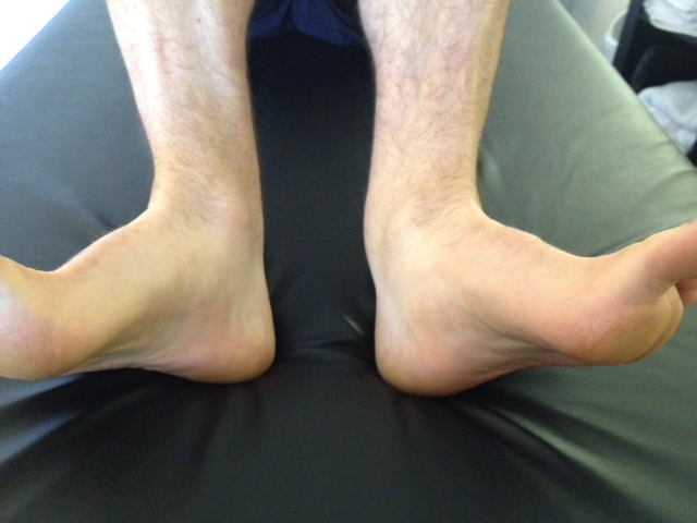
Dr Allen’s post last week on chronic ankle instability (click here for post) served as an inspiration for many of us. It brings to mind the many reasons muscles can become “weak”.

So why does a muscle become weak? We like to categorize the causes as follows:

  • local
  • segmental
  • long loop/cortical

Local causes include muscle injury and muscle pathologies, like muscular dystrophy and neuromuscular endplate disorders like myasthenia gravis. Segmental causes are largely due to reflexes which occur at the spinal cord level. Long loop and cortical causes ae due to an increased inhibition or lack of drive from higher centers, such as the motor cortex and cerebellum.

Lets examine local causes in more detail. To understand causes we must understand what makes a muscle contract.

Muscles are composed of many proteins, 2 of which are actin and myosin (see above). Actin has 2 forms, F (filamental) and G (globular) actin. Imagine 2 grapefruits side by side (G actin) held together in the middle by small filaments (F actin). Now imagine these another set immediately below, in a repeating pattern. These groups of 2 are held together at the sides by an additional protein called tropomyosin. This whole complex looks a little like train tracks. Along the strands of tropomyosin, at regular intervals is yet another protein called troponin. We like to think of troponin as a triangular shaped protein and each part of the triangle has a particular binding site: one for tropomyosin, one for actin and another for calcium ions.

Myosin is another component of muscle, that looks similar to a bunch of golf clubs. The head of the club will, under the right circumstances, interact with actin, the body (tail) of the club interacts with other myosin bodies.

Globular actin and myosin heads are like 2 teenagers and like to interact with one another. Normally, in a resting state, troponin protein covers the active site of myosin binding on G actin. In the presence of calcium, there is a change in shape of the troponin molecule, moving it off of the active site of actin, allowing myosin to bind there. When this happens, the head ratchets and muscle contraction occurs. In the presence of adequate fuel (ie ATP) the myosin head detaches from actin and “recocks”, ready for another contraction cycle (see 2nd picture above).

So where does the calcium come from? It is stored in areas of the muscle called the terminal cisterns. It is released when an action potential fires the peripheral nerve to the neuromuscular endplate of a muscle.

Can calcium be released any other way? Sure it can. How about if the terminal cisterns are damaged, from an injury to the muscle? How about if they are damaged from a disease process?

So, when calcium is released, no matter how it is released, muscles contract. If calcium is not released, then muscles do not contract.

From a local cause, If a muscle is weak, one of the following are usually causing the weakness:

  • there is physical damage to the muscle causing fewer of the working units of the muscle (called sarcomeres) contract

this is by far the most common, due to overuse or trauma

  • there is a problem with the connection of the nerve to the muscle

Disruption of nerve to muscle connections can be also be due to trauma or disease. Weakness that is becoming progressive and worsening, needs to be evaluated further and may be the signal for a progressive muscular or neurological disorder (muscular dystrophy, myasthenia gravis, Gullian Barre, etc)

  • there is insufficient neurotransmitter at the neuromuscular end plate to fire the muscle

this is usually due to a disease process

  • Insufficient calcium could theoretically hamper a muscles contraction, but since calcium is involved with nerve transmission as well, tetany (ie sustained contraction and spasm) would most likely occur due to other reasons that we will not explore at this juncture.

OK, so that sums up local causes. Look for a follow up post about segmental causes next…

We are: The Gait Guys

Last week we ran an archived piece named, “Just because a muscle tests weak doesn’t mean it  needs to be activated”.  Here is the link to that piece.

Today we present some more proof behind our stance on this topic. The referenced article below states in its Context: “An arthrogenic muscle response (AMR) of the soleus and peroneal muscles has been previously demonstrated in individuals with chronic ankle instability (CAI), but the presence of AMR in muscles acting on joints proximal to unstable ankles has not been previously explored.”

And here was their study’s conclusion: “Arthrogenic inhibition of the hamstrings muscles bilaterally and facilitation of the quadriceps muscle ipsilateral to the involved limb were noted in subjects with unilateral CAI. Motoneuron pool excitability appears to be altered in muscles that act on joints proximal to the ankle in those with unilateral CAI.”

This proves our thoughts on a deeper level.  Just because a muscle tests weak does not mean that this muscle is weak or “inactive” or needs  to be “activated”. It is a shame it is not as simple as finding something weak and “activating” it. As we eluded to, this is a far more complex system than that, there are complex feed forward and feedback loops that are mandated and regulated by local cord reflexes, pattern generators and complex cortical loops.  Putting a muscle “back on the grid” prior to activity merely because it tested weak may be putting your client at risk if you are not getting down to the bottom of the problem.  Taking the study mentioned here to a deeper level, finding out that your client has a weak hamstring or quad and negating the possible source of the problem down in an ankle could be devastating to an athlete if they are suddenly returned to aggressive activity immediately after being “activated”.  You may be over riding the central pattern generators, reflex responses and complex cortical loops arthrogenic responses, which could be neuro-protectively calculated. As this article mentioned, “motoneuron pool excitability appears to be altered in muscles that act on joints proximal to the ankle in those with unilateral CAI”.  This is a massively diffuse process going on constantly throughout the body, providing safe mobility and stability. It is not a random process and should not be intervened therapeutically on a random level. At the very least, it requires a patient history, clinical examination, gait evaluation and movement pattern assessment. Anything less is, well, your responsibility if poop hits the fan when they step on the field. 

Shawn and Ivo

Reference:

J Athl Train. 2007 Jul-Sep; 42(3): 355–360.PMCID: PMC1978472Arthrogenic Muscle Response of the Quadriceps and Hamstrings With Chronic Ankle InstabilityEdward J Sedory, MEd, ATC, EMT,Eric D McVey, MEd, ATC,Kevin M Cross, MEd, ATC, PT,Christopher D Ingersoll, PhD, ATC, FACSM, and Jay Hertel, PhD, ATC, FACSM

http://www.ncbi.nlm.nih.gov/pmc/articles/PMC1978472/

“Neural circuits linking activity in anatomically segregated populations of neurons in subcortical structures and the neocortex throughout the human brain regulate complex behaviors such as walking, talking, language comprehension, and other cognitive functions associated with frontal lobes.” 1

We also found this interesting quote from Science Daily on this topic of complex sensory motor behaviors and on the varying information on central pattern generators.

ScienceDaily (June 3, 2012) — “A new finding that motor cortex is a dynamic pattern generator upends existing theory with broad implications for neuroscience.”

“Maybe it is actually easier to understand than we thought. A new paper presents some compelling evidence that the motor cortex, rather than being command central, is more like a part of the machine, sending rhythmic signals down the spinal cord to orchestrate movement.”

"The electrical signal that drives a given movement is therefore an amalgam — a summation — of the rhythms of all the motor neurons firing at a given moment.” This is of course monitored (and modified) by one of our best friends, the cerebellum. 2

The cortex is where movement begins and where it ends; from areas 4, 4s and 6 in the precentral gyrus of the brain’s frontal lobe, down the spinal cord and out to the muscle through the peripheral nerve.   It is also where the information from the body’s receptors feed back,  to give updates on where the body parts are in space (proprioception) and how they are doing functionally (comparing information about length, tension, etc).  It is about sensory and motor function.  Motor function is based on sensory input.  Good motor function is based on good sensory information. It is a subtle, beautiful, intricate symphony.  And when one part goes wrong, the whole system can be thrown off.  

Here is an example we sometimes use in our lectures and with our patients to make this point clear.  Imagine an orchestra playing Beethoven’s beautiful Ode to Joy, a choral symphony for orchestra.  Now imagine one of the musicians begins to play off key. In time, the musicians sitting around that musician who are most locally influenced by that off tune musician, soon become irritated and have troubles playing “in tune”. In time, if not rectified, the whole orchestra could be corrupted and being to take that lead as well.  Hard to believe, but it makes the point that all it takes is one piece not playing well to change the outcome. Similar analogy, all it takes is one weak muscle or one painful joint and the outcome is skewed away from the optimal outcome and in time local dysfunction and compensation becomes an all encompassing compensation. The body’s function and operation, when proper, is an orchestra and orchestration with each piece doing a local job with a more global contribution to the bigger job. When all pieces come together appropriately it creates a symphony of flawless, effortless movement as seen in the video above.

Shawn and Ivo, the gait guys

refs:

1. Front Syst Neurosci. 2014 Feb 13;8:16. eCollection 2014. Cognitive motor interactions of the basal ganglia in development .  Leisman G1, Braun-Benjamin O2, Melillo R3.

tumblr_nn1ca5fx0C1qhko2so1_r1_1280.jpg
tumblr_nn1ca5fx0C1qhko2so2_r1_1280.jpg
tumblr_nn1ca5fx0C1qhko2so3_r1_1280.jpg
tumblr_nn1ca5fx0C1qhko2so4_r1_1280.jpg

Simple Foot Exercises are effective!

Conclusion “These results suggest that the toe spread out (TSO) exercise can be recommended for preventing or correcting HV deformity at an early stage.”

We know and teach that foot exercises work. Here is a nice objective paper (click underlined for abstract) on two exercises we prescribe often.

Here is our variation of the TSO exercise we call the “Lift, Spread and Reach” exercise

Stand comfortably with your feet about shoulder width apart

Stand on your foot tripod with your toes extended. Concentrate on feeling pressure at the center of the calcaneus, the head of the 1st metatarsal and the head of the 5th metatarsal

Lift your toes as high as possible

Spread out (abduct) your toes as much as possible

Reach forward with your toes as far as possible

Place your toes back don on the ground as flat as possible.

repeat 10 X

You can augment the exercise with a rubber band around the toes to provide resistance after you can perform the exercise competently.

Happy exercising!

Ivo and Shawn


Kim MH1, Kwon OY, Kim SH, Jung DY.: Comparison of muscle activities of abductor hallucis and adductor hallucis between the short foot and toe-spread-out exercises in subjects with mild hallux valgus

J Back Musculoskelet Rehabil. 2013;26(2):163-8. doi: 10.3233/BMR-2012-00363.

Gait and any form of locomotion are highly complicated with many pieces necessary to achieve clean, smooth, coordinated motion.
Failure in only one piece of the puzzle can result in profound unhinging of the entire system because of the entangled nature of the feedback loops.
“ Proprioceptive feedback from extensor muscles during the stance phase ensures that the leg does not go into swing when loaded and that the magnitude of extensor activity is adequate for support. Proprioceptive feedback from flexor muscles towards the end of the stance phase facilitates the initiation of the swing phase of walking. Evidence that muscle afferent feedback also contributes to the magnitude and duration of flexor activity during the swing phase has been demonstrated recently. The regulation of the magnitude and duration of extensor and flexor activity during locomotion is mediated by monosynaptic, disynaptic, and polysynaptic muscle afferent pathways in the spinal cord. In addition to allowing for rapid adaptation in motor output during walking, afferent feedback from muscle proprioceptors is also involved in longer-term adaptations in response to changes in the biomechanical or neuromuscular properties of the walking system.” - Lam and Pearson
Neuroscientist. 2004 Jun;10(3):247-59. Cerebellar control of balance and locomotion. Morton SM1, Bastian AJ.

tumblr_nmthzgDyh61qhko2so1_500.jpg
tumblr_nmthzgDyh61qhko2so2_500.jpg

What ischial-femoral impingement might look like as aberrant shoe wear.

A few weeks ago we wrote an article on ischial-femoral impingement. For you to best understand today’s blog post you really should go back and review this interesting clinical phenomenon, here is the link to that piece.

Three weeks ago a talented marathoner came into our office complaining of a long standing deep posterior right hip pain and an equally longstanding left chronic lateral ankle and foot pain.  The ankle had been treated regularly for chronic peroneal tendonitis with various manual therapy modalities and yet the right hip seems to be left out of the equation in terms of treatment.

After taking a detailed history this runner unknowingly pretty much told us they had all the qualifications of ischial-femoral impingement (IFI).  What they did not realize was that they had a cross over gait style that was a significant contributor to the clinical problem.  

Lets now have a look at the shoe wear patterns above. On the left shoe, (a shoe we love, New Balance Fresh Foam (find your next pair at NewBalance Chicago)) we see that the entry zone or crash zone of rear foot impact is heavily worn, especially laterally. Heavy entry zone wear can be from several things, but one thing we always check for and assume until proven otherwise is a cross over gait. It can also just be from excessive rearfoot inversion at foot strike but when excessive there is usually a reason for it, especially when unilaterally as seen here. This foot is not stacking under the knee and hip, it is striking more midline to a plumb line dropped from the hip joint. This creates a steep medial angle of attack. The question is why ? Well, in the history the right hip pain began first and then the left ankle pain, so one should at least consider a compensatory timeline, that being the foot is a compensation in the gait cycle from the painful hip.

This client on examination tested pretty obviously for a right frontal plane drift, meaning when the right foot impacts there is not enough lateral line support to hold the hip/pelvis over the foot and so the pelvis drifts laterally to the right in this case. This can be fought by inverting the foot. This is a strategy to try and stop the lateral drift.  In this case, excessive wear is seen on the entire lateral side of the right shoe to represent this compensation. Changing this clients foot wear, shoe, orthotic is not fixing the problem, in fact it is impairing their ability to compensate and could create more problems, and even another deeper layer of compensation. Again, the inverted/supinated right foot moves the weightbearing line laterally, by moving the foot’s center of pressure from within the confines of the foot tripod towards the lateral border of the foot tripod, in attempt to restack the loading over the laterally drifted hip (hence the right lateral shoe wear pattern). Unfortunately this does not solve the reason for the lateral drifted pelvis. That solution has to come from improved stablization of the hip, pelvis and core and since they tested weak on the right side abdominals, gluteus medius, gluteus max and other  accessory lateral stabilizers,  work must be done there. Interestingly, this runner is stuck into a vicious cycle. The lateral drift to the right is allowing the left hemi-pelvis to dip and this is challenging rotational control of the stance limb and it is causing ischial-femoral impingement (suspecting of the quadratus femoris).  It was clear on examination that there was impairment of the quadratus femoris and obturator externus upon detailed testing and deep palpation was pin point tender over these structures.  Resistance to rotational challenges to the limb, especially iso and eccentric internal rotational challenges, were very poor when it came to coordination, endurance and certainly strength.

Remember, when you are spending time going sideways (right frontal plane drift), you are not spending time moving forwards. This could cause an early right departure and force and early left stance engagement.  But it goes deeper than that in this case.  Here, the right frontally drifting pelvis will pull the left swing leg across the midline with it, creating a left cross over gait.  This will make more sense if you watch our popular video here. Link

So, when this left swing leg is forced into the cross over gait variant, it will force a strong lateral heel strike, as evidenced on the left shoe wear. This is a compensation to what is going on in the right side body mechanics.

Can a cross over occur on one side of the body ? Sure, this case is a perfect example.

Can a cross over gait on the left in this case, cause a vicious cycle and in itself create an environment whereby a right ischio-femoral impingment occurs ?  Sure, neuronal plasticity can be a bitch, it can work in your favor, and against you.

This is not a tough case, if you have seen the beast before and you recognize all of its parameters. If you have not seen the beast before, this case is a nightmare with all these pieces (deep buttock pain, impingement, frontal drift, cross over, strange shoe wear pattern, opposite ankle peroneal pain etc).  Do you have to get this right every time with a bulls eye diagnosis and remedy? Heck no, we flounder every day with new things and variants of old. Sometimes the layers of compensations are so deep that it takes weeks before a recognizable layer presents itself. Patience on both the client and the doctor are necessary.  

So what we have here is a fairly classic shoe wear pattern of a right laterally drifting pelvis and a cross over left leg. In this case it was from a weak right core and pelvis drift creating an environment for the generation of a right ischial-femoral impingement syndrome, driving a left peroneal tendonopathy scenario from the ensuing left cross over gait.  

Remember, don’t fix your clients shoe wear pattern and certainly do not make shoe recommendations from what you see in their shoe wear pattern. Recommending a different shoe to fix this clients problem is a mistake. As is prescribing an orthotic, different foot bed, adding wedges and postings to the shoe or foot bed can also be  mistake. Define the source of the problem, before you go start tinkering around with the bottom of the kinetic chain. Want more ? Try taking our National Shoe fit program to get deeper into this kind of stuff.

We were lucky enough to get this runner’s problem spot on. After many failed attempts by others, this case was 50-75% resolved in one session with the right homework and a great understanding by the runner of their problem. They fully engaged themselves in the understanding of the problem and what they needed to be aware of in their walking and running gait. They were diligent with their homework and understood how it would help the presentation. They presented again to the clinic this week for a focused session to drive the problem further out of town and they are now on their way to the Boston Marathon with a smile and tools to fix the problem. There is a little more fine tuning to do here, but we can wait until they return from Boston.

Good luck in Boston everyone !

We hope this case helps you help someone else, that is the point after all.

Shawn and Ivo, the gait guys

tumblr_nmfyt908iR1qhko2so1_400.jpg
tumblr_nmfyt908iR1qhko2so2_400.jpg

The Serratus and Gait..

Think about the role of the serratus anterior in gait. Now think about it in martial arts. There are profound neuromuscular and fascial connections and implications here. Just like the thoracolumbar fascia which also attaches to the ribs, these muscles seem to be necessary for core stability.

“Conclusions: Simultaneous recruitment of the lower extremity and trunk muscles increases the activation of the SA
muscle during the FPP exercise.
Clinical Relevance: Rehabilitation clinicians should have understanding of the kinetic chain relationships between
the LE, the trunk, and the upper extremity while prescribing exercises. The results of this study may improve clinicians’
ability to integrate the kinetic chain model in a shoulder rehabilitation program. ”

The authors also suggest a hierarchy of exercise to follow. An interesting read for a Sunday.

great full .pdf here: http://www.ncbi.nlm.nih.gov/…/PMC42751…/pdf/ijspt-12-924.pdf

tumblr_nm5tkl3rsl1qhko2so1_1280.gif
tumblr_nm5tkl3rsl1qhko2so2_1280.gif

How well do you understand stance phase mechanics?

Here is a recent question we fielded and thought it would make a great post. 

Question/Comment: I’m slightly confused about closed chain hip motion in the stance leg.


Maybe if I explain what my thought process is you can correct me.  Lets use
left stance phase with the right leg swinging through.

After right mid-swing, the pelvis will be rotating towards the left.  The
motion of the pelvis on the left femur would be relative femur internal
rotation.  I understand that the right leg is externally rotating
(supinating) and that normal open chain kinematics of hip extension is
coupled with external rotation.  But if the pelvis is moving towards the
left AND the left femur externally rotates, wouldn’t that create too much
rotation?  So what I’m saying is that a pelvis that is oriented to the left
with a left femur that externally rotates creates an odd motion in my head
(which may be where the problem lies).  If you’ve ever seen a western where
the gun slingers do that weird walk to a shoot out…that’s what an
externally rotating femur during terminal stance looks like to me.

I’ve discussed this with other clinicians.  Some are in agreement with me,
some think it’s externally rotating, and some don’t know what I’m talking
about.  In my patients I also see a loss of hip IR more than hip ER.  These
patients that lose hip IR seem to have more difficulty in terminal
stance/toe-off phase more than the ones that lose hip ER.

If you could help me understand these kinematics and clear this up for me I
would greatly appreciate it.

Thank you, A

our reply: 

Taking your example with the L leg in stance:
When the L heel contacts the ground, the friction of the ground (hopefully) slow the calcaneus and the talus slide anteriorly on the calcaneus. 

Because of the shape of the calcaneal facets, the talus plantar flexes, adducts and everts. This sets the stage for pronation to occur: the calcaneus everts and the lower leg internally rotates, with the thigh following. The right side of the pelvis is moving to the L (counter clockwise rotation). This should occur (ideally) until midstance. At midstance, the opposite ® foot begins to enter swing phase; this should initiate supination of the stance phase leg (L). At this point, the L foot should be beginning to supinate the the leg and thigh beginning external rotation. It (thigh and leg) should reach maximal external rotation at toe off (maximal counter clockwise rotation of the pelvis) and remain in external rotation until heel strike/initial contact on the L side again. At this point, the pelvis begins clockwise rotation.

It is necessary for the thigh and leg to externally rotate while the pelvis is rotating counter clockwise, because of the constraints of the iliofemoral, pubeofemoral and ishiofemoal ligaments.

We too often see a loss of internal rotation of the hip in symptomatic populations more often than external rotation.

We hope this clarifies things for you.

Thank you again for the question and taking the time to write.

The Gait Guys

The deeper your knowledge and experiences, the more things you will see. As in life, the more experiences you have the wiser you become and the clearer the bigger picture becomes. All these things enrich the experience or observation. These experien…

The deeper your knowledge and experiences, the more things you will see. As in life, the more experiences you have the wiser you become and the clearer the bigger picture becomes. All these things enrich the experience or observation. These experiences take simple black and white and render an infinite palate of grey tones. 

To the untrained observer, these are just two feet. With a little more experience these are two feet of different length. Deeper further, these are two different sized feet with different plantar pressure responses (helped here by increasing the greyscale contrast). Deeper yet, this represents a left foot (viewers right) that has a dysfunctional flexor digitorum longus (FDL) and lateral quadratus plantae muscle. All of these observations allow the skilled and knowledgeable viewer to extrapolate and theorize, with clear thought processes, which leg could be shorter/longer, how the pelvis might be distortioned, step length and stride length variability, foot stability and so much more.

The life long student does not need the contrast enhanced picture on the right to heighten the visibility of the plantar pressures, but it helps.  This is what wisdom and experience do, they enable you to look deeper into something and to see it for what it truly is, not what it appears to be.

Come listen to our teleseminar tonight (Wednesday March 18th, 2015) on www.onlinece.com at 7pm central. Log in early to get set up. Come listen in while we delve into one of the bigger questions, if the left foot (viewers right) is longer it has likely pronated more over a longer period of time stretching out plantar soft tissues and corrupting joint function in multiple areas. But if this is the case, why then are they presenting with plantar pressures that are more representative of supination standards ?  

This is mental gymnastics. It is good stuff to do regularly, even though this is a static presentation, many good theories and thoughts can be brought forth. Getting the answer is not the goal, getting the thought process down is.

The more you know, the more truth you will see.

See you tonight, we will break this down into a microscopic level that will challenge you all.

Shawn and Ivo, the gait guys

The all to common case of the Wobbling Hexapod (Tripod) : Is Your Foot hexa/Tripod Stable Enough to Walk or Run without Injury or Problem ?

Note the music we have chosen today. We tried to match the rate of the dancing tibialis anterior tendon to the tempo of the song, just for fun of course. Well, actually, for neurological reasons as well, as with a steady tempo or beat, your nervous system can learn better. Why do you think we teach kids songs to learn (or you can’t get the theme from the “Jetsons” out of your head).

This is a great video. This client has an obvious problem stabilizing the foot tripod during single leg stance as seen here.  There is also evidence of long term tripod problems by the degree of redness and size (although difficult to see on this plane of view) of the medial metatarsophalangeal (MTP) joint (the MPJ or big knuckle joint) just proximal to the big toe.  This is the area of the METatarsal head, the medial aspect of the foot tripod.

As this client moves slowly from stance into a mild single leg squat knee bend the challenges to the foot’s stability, the tripod, become obvious.  Stability is under duress. There is much frontal plane “Checking” or shifting and the tibial and body mass is rocking back and forth on a microscopic level as evidenced by the dancing tibialis tendon at the ankle level.  The medial foot tripod is loading and unloading multiple times a second. 

Is it any shock to you that this person has chronic foot problems which are exacerbated by running ?  Every time this foot hits the ground the foot is trying to find stability. The medial tripod fails and the big knuckle joint (the 1st MPJ or big toe joint) is enlarging from inflammation, uncontrolled loading through the joint, and early cartilage wear and decay, not to mention the knee falling medially to the foot line as well.  Hallux limitus (turf toe) is subclinical at this time, but it is on the menu for a later date. A dorsal crown of osteophytes (the turf toe ridge on the top of the foot) is developing steadily, soon to block out the range necessary for adequate toe off in this client.  And that means a limitation in  hip extension sometime down the road (and premature heel rise……. did you read Wednesday’s blog post on that topic ?).

*addendum:

Take the time to develop the skill. We ask our clients to work on standing with the toes up to find a clean tripod and do some shallow squats working on holding the tripod quietly. Be sure your glutes are in charge, spin of the limb is in part controlled at the core-hip level so that can a primary location to hunt as well. Eventually work into toes pressed flat but be sure the tripod is still valid, esp the medial tripod. Don’t be what Dr. Allen refers to as a “knuckle popper”. No toe curling/hammering either. Keep that glute on. Move the swing leg forward during a lunge, and then behind you during a squat (mimicking early and late midstance phases of gait/running). This will help your brain realize when it needs this stability and it will also act to press you off balance and will make the foot check and challenge. Do this until you feel the foot fatigue on the bottom. Then Stop. Repeat later. If the medial tripod collapses, the knee will drop inwards and excess pronation is inevitable. We modified this with our prescription of the “100 ups”…..combine the two !

Shawn and Ivo … .  comfortably numb.

Once you have been to the Dark Side of the Moon  (and hopefully you didn’t have any Brain Damage) you will know it well and know what to expect when you return again.  Meaning, when you have seen these issues over and over again, hopefully in your daily work if not regularly here at The Gait Guys, you will quickly know what things to assess and look for in your athletes.  And you might just turn into a Pink Floyd fan at the same time, or at least crave some Figgy Pudding (but you have to eat yer’ meat! How can you have any pudding if you don’t eat  yer’ meat?).

I have been playing with some of this guys work. \
Like respect, flexibility is earned. It is earned through the proper strength, on fundamentally clean motor patterns. Flexibility without strength is not all that common. But we spend so much time stretching and forcing length, and this is a flawed path that will not get you far or to places you truly want to be.  There are many methods, but slow clean movements are important in the early stages, just like in this video. I like to use the 9 second rule in most reteaching in my clinic, 4 seconds to get to the desired end movement, 1 second to hold the end skill, and 4 seconds to get back out of the skill.  Modify as you like and as necessary. Speed can cheat.  Remember, skill first, then endurance and repeatability on the skill, then strength on the endurance based skill.  S.E.S !
This is a Beautiful video, beautiful movements, amazing strength and flexibility.  Things to strive for (and it can’t hurt my Jiu Jitsu either :)  -Dr. Allen

tumblr_nka9cydN3k1qhko2so3_400.jpg
tumblr_nka9cydN3k1qhko2so1_400.jpg
tumblr_nka9cydN3k1qhko2so4_500.jpg
tumblr_nka9cydN3k1qhko2so2_1280.jpg

The partial truth about the Foot Tripod. The HEXApod.

The gait guys have talked about the foot tripod for a very long time. But the truth of the matter is that it is really a HEXApod. HEXA means 6. And when the foot is properly orientated and engaged on the ground, the 5 metatarsal heads and the heel should all be engaged with the ground, truly making it an asymmetrical hexapod. In an ideal scenario, the foot would be most stable if it looked like the strange symmetrical hexapod above with the contact points equally distributed around a center point. But that is not the human foot and this version of a hexapod is far simpler and likely inferior to the foot hexapod when human locomotion is to be attempted. The human foot is engineering marvel when it works properly.  

Perhaps the best example of what I mean by the foot being a HEXApod is in the pressure diagram above. In that first picture, on the right of that picture, we see multiple pressure points under the metatarsal heads of the right foot.  Minus the missing 1st metatarsal head pressure point (taken over by increased flexor hallucis longus activity represented by increased pressure at the big toe),  this pretty much confirms that the foot is not a tripod, rather a hexapod. The theory of the tripod, the 1st and 5th metatarsal heads and the heel, is only crudely accurate and honest. In this picture case, this person has increased lateral foot weight bearing (possibly why the 1st MET head pressure is absent) and possibly represented by pressure under the base of the 5 metatarsal. This is not normal for most people and if this person could get the 1st MET head down, they might even have a HEPTApod foot structure because of the 5th metatarsal base presentation (which sometimes represents peroneal muscle weakness). 

Where did we lead you astray after all these years of “tripod” talk ? We have always discussed the foot tripod. We have always discussed the imperative need to keep the limb’s plumb line forces within the area represented by the tripod.  If your forces fall more laterally within the tripod, as in this first discussed picture, one is at increased risk of inversion events and the myriad of compensations within the entire body that will occur to prevent that inversion. So again, why the tripod?  Well, it is easier to understand and it serves our clients well when it comes to finding active foot arch restoration as seen in this video of ours here.  But, the truth of the matter is that all of the metatarsal heads should be on the ground. The 2nd METatarsal is longer, the 3rd a little shorter, and the 4th and 5th even a little short than those. With the 1st MET shorter, these 5 form a kind of parabolic arc if you will. Each metatarsal head still should contact the ground and then each of those metatarsals should be further supported/anchored by their digits (toes) distally.  So the foot is actually more truly a HEXAPOD. Take the old TRIPOD theory we have always spoken about and extend a curved line beyond the forefoot bipod points (1st and 5th metatarsals) to incorporate contact points on the 2, 3 and 4th metatarsal heads. These metatarsals help to form the TRANSVERSE arch of the foot. It is this transverse arch that has given us the easily explainable foot TRIPOD because if a line is drawn between just the shorter 1st and 5th metatarsals, we do not see contact of the 2-4 metatarsal heads when we only look for pressure between these two bipod landmarks, but the obvious truth is that the 2-4 metatarsals are just longer and extend to the ground further out beyond this theoretical line drawn between the 1st and 5th MET heads.   

So, the foot is a HEXAPOD. Make no mistake about it. It is more stable than a tripod because there are more contact points inside the traditionally discussed foot tripod model. And frankly, the tripod theory is just a lie and just too fundamentally simple, unless you are an American 3 toed woodpecker.

Dr. Shawn Allen,     www.doctorallen.co

one of the gait guys

Some stuff you need to know about running spikes.I see many track runners in my office, from middle school all the way into the USA Masters Division.  A few years ago one of the top USA Masters Milers came to see me on Friday before heading off to a…

Some stuff you need to know about running spikes.

I see many track runners in my office, from middle school all the way into the USA Masters Division.  A few years ago one of the top USA Masters Milers came to see me on Friday before heading off to a national meet. He showed me some of his spikes (see pics above) and complained the there was something off on the spikes on the left, the Nike Mambas.  The shoe to the right is the Nike Zoom Miler.

You need to understand a bit of the physics of running turns to understand what is missing for this runner in this pair of spikes.  Things do change if you are running on a sloped track, but those are only found indoors and are not all that common to run on for most folks so we will stick with the thinking on flat tracks.

What you should be able to easily detect is that the Nike Mamba’s are missing the lateral 5th metatarsal forefoot spike on the cleat plate.  And you need to then realize that this is the right shoe, so it is the outside foot/leg on the track. It is the foot that will be pushing off harder from the outside on the turns to keep the centripetal forces of running a curve from allowing the runner to fall off the curve into the outer lanes. This right foot will always be pushing from outside to inside to maintain the body’s progression in the desired lane, when running the curves.

Think about it for a minute. In order to run in a circle, or a curve in this situation, the outside foot always has the tendancy to be more inverted to keep foot contact on the ground. This is where a Forefoot varus MIGHT come in handy ! This means the foot will be tipped to the outside a little, because of the curve and because the body will be leaning into the center of the track on the curves. Thus the foot and shoe will be relying on more lateral foot pressures to drive the body mass back into the lane since centripetal forces will always be driving you laterally out of the lane.  Thus, the lateral spikes on the right foot must be accommodating.  In the case of the Mamba shoe. there is only a sheet of black hard plastic over the midshaft-head of the 5th Metatarsal on the lateral foot. It is no wonder the runner was feeling like he was slipping on the turns (the front of the midfoot was not anchored to the ground, only the forefoot due to the spikes in that location). You can see clear evidence of the lateral slipping in the picture. Can you see the orange/brown patch where he was slipping ? A spike there in that area would have been wonderful.  Slipping is a power leak and a risk for injury.  If the foot is trying to gain purchase into/onto the track with the foot inverted there needs to be traction at that lateral foot, what is referred to as the Lateral Column.  You can see why the Nike Zoom Miler was a better choice, there is a nice spike placement under the lateral foot for just this measure, and there is no evidence of slippage wear.  He told me that the Mamba was a steeplechase designed shoe but we still both felt that the issue remained relevant even in that event. The Nike website however states that “the Nike Zoom Mamba Men’s Track and Field Shoe is perfect for the 800-5000m track athlete” so we think they have missed an issue here in our opinion.

I could make a better case for the Mambas if  they were for a 100m straight run but I would still like a 5th metatarsal /lateral spike where there isn’t one.  I will occasionally file spikes to get the perfect feel for the athlete.  It is usually the 5th metatarsal and 1st metatarsal spikes I mess with, merely to help hone the athletes feel on the track. The problem is that each track has a different feel so it is less of an occurrence in recent years.

It is good to know your shoes, it is good to know your physics. It is great to know them both and melt them together to solve problems.  Not all spikes are created equal, not all tracks are the same, not all events are the same and certainly not all feet and the athlete’s who own them are the same.  And on the topic of Forefoot Foot types, both the forefoot varus and forefoot valgus foot might have a problem with the Mamba’s depending on their strength, skill and strategies for ground purchase.  Hopefully your shoe store and your track and cross country coaches know these issues. You might want to bring this blog post to their attention however, just in case.

Dr. Shawn Allen

Tom Purvis hits some strong points in this video about squatting, hip hinges, ankle dorsiflexion, and movement as a whole.  * Keep in mind, this is all sagittal plane stuff….. it gets far more complicated when there are lateral (frontal) plane or rotational (axial) considerations ….. these are the “knees out” dialogues and debates you have read over and over on the web in the last year.

Dr. Shawn Allen

addendum:

Food for thought after posting today’s Tom Purvis squat video.

Could this study below translate into the statement/question: 

“attempting to achieve sufficient dorsiflexion through the combined ‘foot pronation-ankle dorsiflexion’ mechanism, as opposed to just dorsiflexion from the ankle mortise joint alone, may change the dynamics of the entire limb…. in this case, hip flexion range observation. Is this because when dorsiflexion is cheated via foot pronation, instead of just ankle dorsiflexion, there is more internal tibia/femoral spin than would normally occur from just sagittal ankle hinging which can in turn impair terminal hip flexion range via impingement type action ? I think so. It would be cool to see what would have happened in the study had the pronating clients been shown my foot tripod restoration exercise (it’s on youtube).   -Dr. Allen

here is some new research on this point, for what it is worth.  It keeps the mind thinking though.

J Phys Ther Sci.  2015 Jan;27(1):285-7. doi: 10.1589/jpts.27.285. Epub 2015 Jan 9.The kinematics of the lower leg in the sagittal plane during downward squatting in persons with pronated feet.  Lee,Koh da,  Kim 

Abstract

[Purpose] This study aimed to examine changes in lower extremity kinematics in the sagittal plane during downward squatting by subjects with pronated feet. [Subjects and Methods] This study selected 10 subjects each with normal and pronated feet using a navicular drop test. The subjects performed downward squatting, in which the knee joints flex 90° in a standing position. We recorded the angles of the hip, knee, and ankle joint in the sagittal plane through motion analysis. For the analysis, the squatting phase was divided into phase 1 (initial squat), phase 2 (middle squat), and phase 3 (terminal squat) according to the timing of downward squatting. [Results] In the pronated foot group comparison with the normal group, thehip joint flexion angle decreased significantly in phases 2 and 3. The dorsiflexion angle of the ankle joint increased significantly in phase 3. The flexion angle of the knee joint did not differ between groups in any of the phases. [Conclusion] The pronated foot group utilized a different squat movement strategy from that of the normal foot group in the sagittal plane.