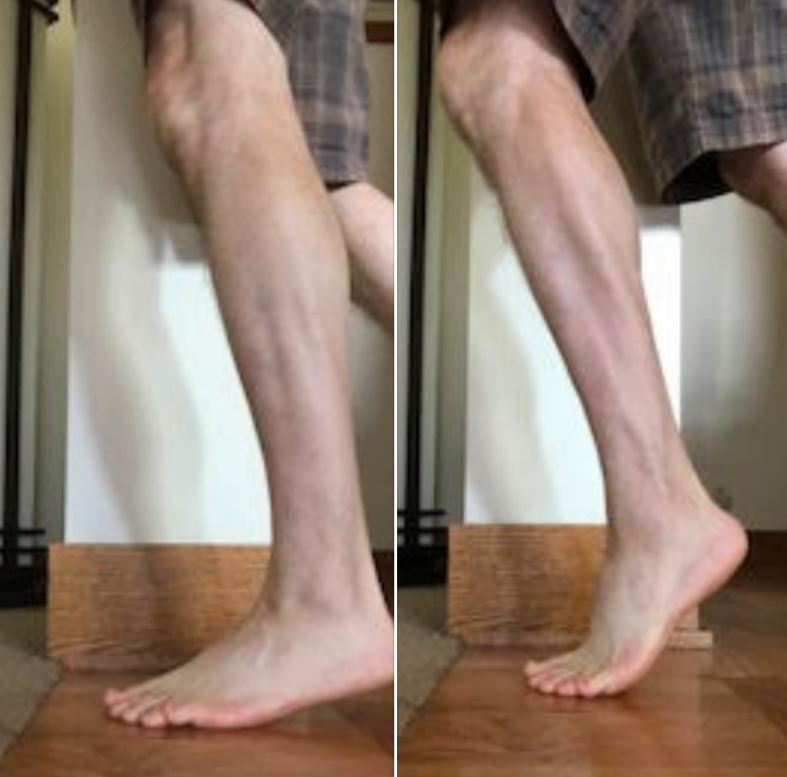
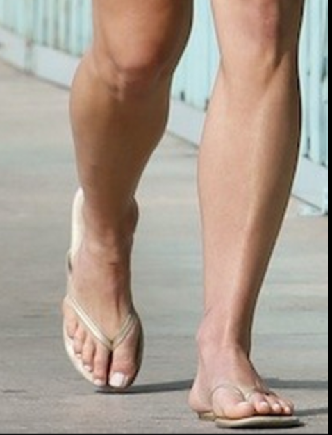
Forefoot running, achilles loads & gait retraining

tag/key words: gait, gaitproblems, gaitanalysis, forefootrunning, forefootstrike, achilles, heelstrike, elastography, thegaitguys, microvascularity, rockeredshoes, HOKA, metarocker, gaitretraining,

Links to find the podcast:
Look for us on iTunes, Google Play, Podbean, PlayerFM and more.
Just Google "the gait guys podcast".

Our Websites:
www.thegaitguys.com
Find Exclusive content at: https://www.patreon.com/thegaitguys
doctorallen.co
summitchiroandrehab.com
shawnallen.net

Our website is all you need to remember. Everything you want, need and wish for is right there on the site.
Interested in our stuff ? Want to buy some of our lectures or our National Shoe Fit program? Click here (thegaitguys.com or thegaitguys.tumblr.com) and you will come to our websites. In the tabs, you will find tabs for STORE, SEMINARS, BOOK etc. We also lecture every 3rd Wednesday of the month on onlineCE.com. We have an extensive catalogued library of our courses there, you can take them any time for a nominal fee (~$20).

Our podcast is on iTunes and just about every other podcast harbor site, just google "the gait guys podcast", you will find us.

Where to find us, the podcast Links:

iTunes page:
https://itunes.apple.com/us/podcast/the-gait-guys-podcast/id559864138?mt=2

Google Play:
https://play.google.com/music/m/Icdfyphojzy3drj2tsxaxuadiue?t=The_Gait_Guys_Podcast

Direct download URL: http://traffic.libsyn.com/thegaitguys/pod_150.2_-_42719_5.00_PM.mp3

Permalink URL: http://thegaitguys.libsyn.com/forefoot-running-achilles-loads-gait-retraining

Libsyn Directory URL: http://directory.libsyn.com/episode/index/id/9555122

Show notes:

Ultrasound elastographic assessment of plantar fascia in runners using rearfoot strike and forefoot strike. Tony Lin-WeiChen et al
https://www.sciencedirect.com/science/article/pii/S0021929019302775
J Sci Med Sport. 2015 Mar;18(2):133-8. doi: 10.1016/j.jsams.2014.02.008. Epub 2014 Feb 14.

Rocker shoes reduce Achilles tendon load in running and walking in patients with chronic Achilles tendinopathy.. Sobhani S et al
https://www.ncbi.nlm.nih.gov/pubmed/24636129/


The increase in muscle force after 4 weeks of strength training is mediated by adaptations in motor unit recruitment and rate coding. Alessandro Del Vecchio et al
https://physoc.onlinelibrary.wiley.com/doi/10.1113/JP277250


Learning new gait patterns is enhanced by specificity of training rather than progression of task difficulty. ChandramouliKrishnan et al
https://www.sciencedirect.com/science/article/pii/S0021929019301927


The microvascular volume of the Achilles tendon is increased in patients with tendinopathy at rest and after a 1-hour treadmill run. Pingel J et al
Am J Sports Med. 2013 Oct;41(10):2400-8. doi: 10.1177/0363546513498988. Epub 2013 Aug 12.
https://www.ncbi.nlm.nih.gov/pubmed/23940204/

*** Our PODcast disclaimer:
This podcast is for general informational purposes only. It does not constitute the practice of medicine, nursing, rehab, treatment, therapy recommendations or anything of the sort. This podcast should not replace proper medical advise that should only be attained through proper medical channels that would entail a full medical and/or biomechanical physical examination and/or appropriate diagnostic testing. No doctor-patient relationship is formed by listening to this podcast or any information gleaned from our writings or social media work.
The use of this information and the materials linked to the podcast is taken at the users own risk. This podcast and the content shared is not intended to replace or be a substitute for appropriate professional medical advise diagnosis or treatment. Users should not disregard or delay obtaining medical advice for any condition they have and should seek the advice and assistance from their providers for any such conditions.

Top end heel raises. The top end might matter.

Screen Shot 2019-04-19 at 6.34.09 PM.png

Thought experiment . . .
If top end posterior compartment (loosely, the calf complex) strength is lacking, then heel rise may not be optimized to transfer body mass forward sufficiently and effectively.
This lack of forward progression, fails to move the body mass sufficiently forward enough to reduce the external moment arms and optimize the internal moment arms to take maximal advantage of the calf complex (I am talking about moment arms between the grounded 1st MTP joint and ankle mortise & ankle mortise and achilles tendon).
These are rough thoughts today gang, letting you inside our heads and how we juggle multiple parameters when we are struggling to solve a client's problems.

In the lower heel rise photo, The body mass does not progress forward enough over the grounded first MTP joint at the big toe (during gait, the heel doesn’t just rise up, the axis of the ankle joint moves both up and forward).
In this case, the foot may not be fully rigid in a supinated position to benefit from joint closed-packed positions. Thus, the foot may be more pliable and one might suppose that if not adequately supinated, they are inadequately still too much relatively pronated. This might put more load into the tibialis posterior and other soft tissue mechanical loading scenarios that are less optimally suited to do this job. Over time, might this lead to pathology? Likely.
Thus, when running on a weaker posterior mechanism (often found unilaterally) the higher up posterior chains might be overburdened, the tendon loads and loading response of the achilles, tibialis posterior, and long flexors will be most likely altered, likely negatively, the naturally occurring foot locking mechanisms might be less optimal than desired, subtalar and forefoot loading might be premature (ie. sesamoid malpositioning for one, as a simple example), etc etc. Loading a foot(the mid and forefoot) into heel rise that is still somewhat pronated creates a different moment arm around the subtalar joint axis (that moves through the 1st metatarsal), than a foot that is more supinated.

Now, put these ideas into the 2 photos from yesterday where one might be loading the forefoot laterally or more medially, and now make the top end strength more in one of those scenarios. Is it any wonder why so many struggle with posteiror mechanism tendonopathies ? There are so many parameters to consider and examine. And, if not examined in great detail, the key lacking parameter can be missed.
Hence, just forcing calf strength loading is too simple a solution, there is a needle in that haystack that upset the client's apple cart, it is the job of the clinician to find it and remedy it.

Today, looking into the research and finding some interesting things that are spurring some thoughts.

Shawn Allen, one of the gait guys

You are mostly likely not getting to your big toe at push-off if you are doing this.

Screen Shot 2019-04-08 at 4.20.41 PM.png

You are mostly likely not getting to your big toe at push-off if you are doing this. And, if you have a painful big toe, you will do it as well. Oh, and Head-over-foot related, yup. Read on . . .


Yes, the cross over gait. Yes, when you are into a cross over gait you are most certainly head over foot. And that is most likely not a good thing.


If you are not closer to stacking the hip over the knee, and knee over the foot (like in the photo "SUI" bib runner) you are not likely getting to much of your big toe at terminal stance loading, when you could be getting more power at push off.
Said another way, if you are attacking the ground with the feet closer together, as if you are running on a line (as in the photo) you are going to be more on the outside of the foot (note the lateral foot contact), show a similar wear/loading pattern as in these shoes, and hardly load the medial foot tripod effectively.
Go ahead, walk around your office or home right now . . . . with a very narrow step width and see how little you can load into the bit toe-medial foot tripod (note how little effective glute engagement you get as well). Then, walk with a wider step width, note the easier more effective big toe-medial tripod loading, and, not the glutes come into play.

Screen Shot 2019-04-08 at 4.20.52 PM.png

Thus, head over foot/cross over gait is foolish for effective gait. You have a big toe, don't you wish to use it ? One has to find that balance between an economical step width that still allows an effective toe off event in walking and running. A very narrow cross over-style gait does not afford us this.
So, should it be any surprise to any of us that someone with pain in the big toe or medial tripod complex will choose a narrow step width to avoid the painful loading ? No, no surprise there at all.
We have been writing about the cross over gait for 10 years, bringing little pieces of research to the forefront to prove our theories on it as the research presents itself. We first brought it to you with our 3 part video series here. Search our blog, type in "cross over gait" into the search box on the site www.thegaitguys.com and get a LARGE coffee before hand, you are going to be reading for several hours.

https://www.youtube.com/watch?v=LG-xLi2m5Rc
https://www.youtube.com/watch?v=WptxNrj2gCo
https://www.youtube.com/watch?v=oJ6ewQ8YUAA

Screen Shot 2019-04-08 at 4.21.31 PM.png
Screen Shot 2019-04-08 at 4.21.23 PM.png

Support for visual gait analysis... with respect to leg length discrepancies


image credit: https://pixabay.com/photos/eye-blue-eyelashes-vision-make-up-691269/

image credit: https://pixabay.com/photos/eye-blue-eyelashes-vision-make-up-691269/

We talk about leg length discrepancies all the time here on the blog and sometimes, how small discrepancies cause changes in peoples biomechanics. The gold standard for measuring a leg length difference is full length lower extremity X ray, but this presents a problem due to the ionizing radiation, accessibility as well as impracticality of X rays every person with a suspected difference.

We have talked about different compensations as to how to get around a leg length discrepancies. Last week we actually did a tell a seminar on this entire subject. Your patient or client needs to “create clearance” for the longer leg side. This can be accomplished in many ways.

The 5 most common strategies (keep in mind there are many more) are:

  • lean the torso to the short leg side (essentially hip adduction of the longer side)

  • hike the torso on the long leg side

  • circumduct the longer lower extremity

  • increase plantar flexion of the calf of the short leg side

  • increase hip and knee flexion on the longer leg side

And that is exactly what this study found. They looked at kinematics in people with anatomical leg length discrepancies and found that hip adduction as well as increased hip and knee flexion were 2 variables that were consistent in folks with anatomical differences and suggest these variables are a useful screening tool.

Paying attention to how people move and looking for asymmetries. In our opinion, that’s the name of the game : )

Dr Ivo Waerlop, on of The Gait Guys

Zeitoune GNadal JBatista LAMetsavaht LMoraes APLeporace G.Prediction of mild anatomical leg length discrepancy based on gait kinematics and linear regression model. Gait Posture. 2019 Jan;67:117-121. doi: 10.1016/j.gaitpost.2018.09.027. Epub 2018 Sep 29.

#LLD #leglengthdifference #leglengthinequality #visualgaitanalysis #thegaitguys #gaitanalysis

Pronation anyone? Coupling? Walking Speed? How do they relate?

We have talked many times here on TGG about pronation, supination, overpronation, asymmetrical pronation, and more.

When most people think of pronation, they think of midfoot pronation, or pronation about the subtalar or transverse tarsal joints. Pronation can actually occur about any articulation or bone, but with respect to the foot, we like to think of rearfoot (ie. talo-calcaneal), midfoot (talo-navicular) and forefoot (transverse tarsal). The question is why does this matter?

Pronation, with respect to the foot, is defined as a combination of eversion, abduction and dorsiflexion (see picture attached) which results in flattening of the planter vault encompassing the medial and lateral longitudinal arches. In a normal gait cycle, this begins at initial contact (heel strike) and terminates at midstance, lasting no more than 25% of the gait cycle.

In a perfect biomechanical world, shortly following initial contact with the ground, the calcaneus should evert 4-8 degrees, largely because the body of the calcaneus is lateral to the longitudinal axis of the tibia. This results in plantar flexion, adduction and eversion of the talus on the calcaneus, as it slides anteriorly. At this point, there should be dorsiflexion of the transverse tarsal (calcaneo-cuboid and talo-navicular joints). Due to the tight fit of the ankle mortise and its unique shape, the tibial rotates internally (medially). This translates up the kinetic chain and causes internal rotation of the femur, which causes subsequent nutation of the pelvis and extension of the lumbar spine. This should occur in the lower kinetic chain through the 1st half of stance phase. The sequence should reverse after the midpoint of midstance, causing supination and creating a rigid lever for forward propulsion.

Pronation, along with knee and hip flexion, allow for shock absorption during throughout the 1st half of stance phase. Pronation allows for the calcaneo-cuboid and talo-navicular joint axes to be parallel making the foot into a mobile adaptor so it can contour to irregular surfaces, like our hunter gatherer forefathers used to walk on before we paved the planet. Problems arise when the foot either under pronates (7 degrees valgus results in internal tibial rotation), resulting in poor shock absorption or over pronates (> 8 degrees or remains in pronation for greater than 50% of stance phase).

This paper talks about how foot and ankle pathologies have effects on other articulations in the foot. They looked at stance phase of gait in 14 people without pathology at 3 different walking speeds. they found:

coupling relationships between rear foot inversion and hallux plantar flexion and rear foot eversion with hallux dorsiflexion.... When the rear foot everts (as it does as discussed above) during pronation from initial contact to mid stance , the hallux should be extending AND when the rear foot everts, as it should from mid stance to terminal stance/pre swing, the hallux should be plantar flexing to get the 1st ray down to the ground

medial (internal) rotation of there leg was accompanied by mid foot collapse (read pronation) and lateral (external) rotation with mid foot elevation (read supination).... Because of the shape of the talar dome and shape of the talo calcaneal facet joints, the talus plantar flexes, everts and adducts from initial contact to mid stance, and dorsiflexes, inverts and adducts from mid stance to terminal stance/ pre swing

walking speed significantly influenced these coupling relationships....meaning that the faster we go, the faster these things must happen and the greater degree that the surrounding musculature and associated cortical control mechanisms must act

So, when these relationships are compromised, problems (or more often, compensations) ensue. Think about these relationships and the kinetics and kinematics the next time you are studying someones gait.

Dr Ivo Waerlop, one of The Gait Guys

Dubbeldam R1, Nester C, Nene AV, Hermens HJ, Buurke JH. Kinematic coupling relationships exist between non-adjacent segments of the foot and ankle of healthy subjects.Kinematic coupling relationships exist between non-adjacent segments of the foot and ankle of healthy subjects.Gait Posture. 2013 Feb;37(2):159-64. doi: 10.1016/j.gaitpost.2012.06.033. Epub 2012 Aug 27

cool video on our blog to go with this post:

https://www.thegaitguys.com/thedailyblog/2018/8/7/what-do-you-know-about-pronation-and-supination

#gaitanalysis #thegaitguys #pronation #couplingrelationshipsandgait #pronation

Case Studies in Gait Analysis: Focus on the Short Leg (online video class)

Case Studies in Gait Analysis: Focus on the Short Leg
*link is below

*this is the online Continuing education class we did last week, for those of you who could not get to the Wednesday evening class.
*our entire catalogue of lectures and seminars are all here on this site for CE/CEU

Case Studies in Gait Analysis: Focus on the Short Leg
- Review anatomical vs functional short leg
-Review the kinematics and kinetics of the short leg during the gait cycle
-View and discuss case studies looking at functional and anatomical short legs
-Predict pathomechanics that will arise from a short leg
-Propose remedies for the gait abnormalities seen

Link: https://chirocredit.com/course/Chiropractic_Doctor/Biomechanics_211

Screen Shot 2019-04-22 at 1.20.16 PM.png

The hip flexors do not pull the leg forward during swing (mostly).

The hip flexors are not responsible for pulling/flexing the swing leg forward in gait or running. The psoas is a mere swing phase perpetuator, not an initiator.
For about 2 decades we have been saying in our lectures, posts and podcasts that it is the reduction of the obliquity of the pelvis during gait from various other tissues and biomechanical events that causes leg swing, meaning the trail leg is brought forward in swing largely by the abdominal muscle linkage to the pelvis (and other loaded tissues) that is responsible for forward swing of the leg. It is not the hip flexor group that does this hip flexion action. Thus it could be considered foolish to train the hip flexors to be the primary swing drivers. Here is another supporting piece of research.

"These experiments also showed that the trailing leg is brought forward during the swing phase without activity in the flexor muscles about the hip joint. This was verified by the absence of EMG activity in the iliacus muscle measured by intramuscular wire electrodes. Instead the strong ligaments restricting hip joint extension are stretched during the first half of the swing phase thereby storing elastic energy, which is released during the last half of the stance phase and accelerating the leg into the swing phase. This is considered an important energy conserving feature of human walking. "

Dan Med J. 2014 Apr;61(4):B4823.
Contributions to the understanding of gait control.
Simonsen EB1.

More on the scourge of Flip Flops. Riding the inside edge of the sandal. Mystery hunting with Dr. Allen.

Screen Shot 2019-04-19 at 7.53.35 PM.png

Tis the season upon us. Riding the inside edge of the sandal.
You can see it in the photo, the heel is a third of the way off the sandal.

You either have it or have seen it. It is frustrating as hell if you have it. Your heel rides on only half of your flip flop or summer sandals. You do not notice it in shoes, only in sandals, typically ones without a back or back strap. This is because the heel has no controlling factors to keep it confined on the rear of the sandal sole. There is no heel counter on open backed shoes and sandals, the counter keeps the heel central on the back of the footwear. There is a reason this inside edge riding happens to some, but not everyone. It is best you read on, this isn’t as simple as it might seem.

These clients may have restricted ankle rocker (dorsiflexion), restricted hip extension and/or adductor twist (if your reference is the direction the heel is moving towards). I could even make a biomechanical case that a hallux limitus could result in the same scenario. So what happens is that as the heel lifts and adducts it does not rise directly vertically off the sandal, it spins off medially from the “adductor twist” event. This event is largely from a torque effect on the limb from the impaired sagittal mechanics as described above, manifesting at the moment of premature heel rise resulting in an slightly externally rotating limb (adducting heel). The sandal eventually departs the ground after the heel has risen, but the sandal will rise posturing slightly more laterally ( you can clearly see this on the swing leg foot in the air, the sandal remains laterally postured). Thus, on the very next step, the sandal is not entirely reoriented with its rear foot under the heel, and the event repeats itself. The sandal is slightly more lateral at the rear foot, but to the wearer, we believe it is our heel that is more medial because that is the way it appears on the rear of the sandal or flip flop. Optical illusion, kind of… . . a resultant biomechanical illusion is more like it.

Screen Shot 2019-04-19 at 7.53.46 PM.png


You will also see this one all over the map during the winter months in teenagers who swear by their Uggs and other similar footwear, as you can see in the 2nd photo above. This is not an Ugg or flip flop problem though, this is often a biomechanical foot challenge that is not met by a supportive heel counter and may be a product of excessive rear foot eversion as well. This does not translate to a “stable” enough shoe or boot, that is not what this is about. This is about a rearfoot that moves to its biomechanical happy place as a result of poor or unclean limb and foot biomechanics and because the foot wear does not have a firm stable and controlling heel counter. This is not about too much pronation, so do not make that mistake. And orthotic is not the answer. A heel counter is the answer. The heel counter has several functions, it grabs the heel during heel rise so that the shoe goes with the foot, it give the everting rearfoot/heel something to press against, and as we have suggested today, it helps to keep the rearfoot centered over the shoe platform. To be clear however, the necessary overuse and gripping of the long toe flexors to keep flip flops and backless sandals on our feet during the late stance and swing phases of gait, clearly magnifies these biomechanical aberrations that bring on the “half heel on, half heel off” syndrome.

There you have it. Another solution to a mystery in life that plagues millions of folks.

Shawn Allen, the other gait guy

So forget repairing your ACL tear huh?

Soapbox rant today: So forget repairing your ACL tear huh?

Just give it some deep thought before you decide rehab is enough for you. Don't get fully sucked into the non-surgery hype, sometimes there is value and purpose. We are not necessarily saying that we are pro-ACL surgery, but it does have a place when we are talking about a major ligament with many functions beyond articular vector restraint.

*Here is where we see the present problem with the "newer" rehab-only hype for ACL tears . . . . the follow up time frames of the research pieces that suggest that ACLR is sufficient, in our opinion are not long enough into the future (years) to substantiate that secondary instability is not occurring or not a risk. In fact, there are enough articles to substantiate that secondary instability (often deeply rotational) will occur if no ACL repair occurs.

But, other bad things can happen if the joint is not cinched up tightly.
"Increases in TFI (time from injury) are associated with medial meniscal tears, including irreparable medial meniscal tears, medial femoral condyle chondral damage, and early medial tibiofemoral compartment degenerative changes at time of ACLR. These findings highlight the importance of establishing a timely diagnosis and implementing an appropriate treatment plan for patients with ACL injuries. This approach may prevent further instability episodes that place patients at risk of sustaining additional intra-articular injuries in the affected knee. "
*in this study 47.2% were classified as playing competitive or professional sports versus recreational sport

There have been some therapists in the field around the world that have been promoting that ACL surgeries ** are seemingly becoming more and more unnecessary. Their stance seems to be that with hardcore rehab that the knees do just as well, that performance is not lost. Sure, this is possible this or next season, but what about in 2 years? 5 years ? And what will the consequences be then? This article outlines some thoughts.
So, lets just all be careful of the strong points of view we put out there for the consumer. We get their point, but it is foolish to dismiss that the ligament doesn't have a function and is never necessary to replace/repair as this article (and many others report). SECONDARY instability is a real thing, rotational instability in non-ACL repaired** knees is a real thing. Attenuation of secondary joint restraints over time is a real thing, and the cost that comes with those changes. The consequences to the joint structure as secondary instability sneaks in, are a real thing, they are most likely to occur, even if you rehab your client's knee deeply. So be sure that you educate your client, that without their ACL their knee will never be as good, even if you are a champion rehab guru, you are just not that good that you and your rehab can negate all of the rotational vectors of loading in your high level athletes. Time and load will win, just be honest. Just because you do not see consequences tomorrow, just because your top-tier athlete continues to perform this season at top levels without compliant, doesn't mean they will not be present next year. Just be up front with your clients.
And here is another thought to chew on. 24 months ago my Jui-jitsu master Prof Carlos Lemos Jr. tore his ACL. We rehabed and he did well, he even won his 4th world championship without his ACL. But, we had these talks, and he knew that even though he was able to perform at the top level, he knew that the leg was not like the other. He decided 6 weeks ago to have it repaired because we discussed many times the above kinds of long term possibilities. I placed what facts and experiences I have had over 20+ years, the research that is presently out there, and let him decide. He decided that "hope" only goes so far, that he knows he will not be exceptionally as strong on the long term rehab to the degree it was initially performed, and he did not want to risk subsequent internal joint damage that might ensue.
Yes, not everyone needs ACL surgery, especially those who are not highly active or sporting, or the aging/elderly, but we can make a case that just rehabing and dismissing repair is also going to miss some vital points. We are not saying that we are pro-ACL surgery, but it does have a place.
Just educate your client honestly, then let them decide the direction, and do good work.

If anyone wishes to debate here, lets do it. But come at us with 5-10 year post-rehab no-ACL surgery cases with MRI's showing no intra-articular cost. (Good luck with that.) But if you find such unicorns, we definitely want to see them so we can share it and adjust our stance more softly. We want to be as smart and accurate on our rants as possible, it is important.

**corrected/ammended 10:57central time

photo credit: pixabay.com. thank you !

Orthop J Sports Med. 2018 Dec 11;6(12):2325967118813917.
Relationship Between Time to ACL Reconstruction and Presence of Adverse Changes in the Knee at the Time of Reconstruction.
Sommerfeldt M1,2, Goodine T2, Raheem A3, Whittaker J1,4, Otto D

How do humans run faster ?


Runners appear to use two different strategies to increase their speed according to this article we are appreciating by Dorn, Schache and Pandy


In specific observance of the lower limb muscles,
"Humans run faster by increasing a combination of stride length and stride frequency. In slow and medium-paced running, stride length is increased by exerting larger support forces during ground contact, whereas in fast running and sprinting, stride frequency is increased by swinging the legs more rapidly through the air". . . . .
"For speeds up to 7ms–1, the ankle plantarflexors, soleus and gastrocnemius, contributed most significantly to vertical support forces and hence increases in stride length. At speeds greater than 7ms–1, these muscles shortened at relatively high velocities and had less time to generate the forces needed for support. Thus, above 7ms–1, the strategy used to increase running speed shifted to the goal of increasing stride frequency. The hip muscles, primarily the iliopsoas, gluteus maximus and hamstrings, achieved this goal by accelerating the hip and knee joints more vigorously during swing."

Muscular strategy shift in human running: dependence of running speed on hip and ankle muscle performance
Tim W. Dorn, Anthony G. Schache and Marcus G. Pandy*
Department of Mechanical Engineering, University of Melbourne, Victoria 3010, Australia. The Journal of Experimental Biology 215, 1944-1956
© 2012. Published by The Company of Biologists Ltd
doi:10.1242/jeb.064527

More on tendon stiffness

There has been much discussion about tendon stiffness in the last few years. Here is an interesting paper.

Here is a piece of the authors' hypothesis. " If tendons can be overloaded, their mechanical properties should change during exercise. "
The present study measured AT stiffness before and after a marathon.
Here is what they found in this study, "AT stiffness did not change significantly from the pre-race value of 197±62 N mm−1 (mean ± s.d.) to the post-race value of 206±59 N mm−1 (N=12, P=0.312). Oxygen consumption increased after the race by 7±10% (P<0.05) ". This was a small N study, but that leaves room for more large scale studies to see if it holds up.
What remains interesting and head scratching to us is that a tendon has its tension developed by the muscle contracting that is attached to it. So, one would think that a marathon would cause some fatigue in the calf which would change the tension in the achilles. But we are brought to the thought that perhaps stiffness and tension are not the same animals, not even close ?
However, the article mentions this, "A typical training effect, regardless of whether training is plyometric or isometric resistance training, is an increase in AT stiffness (Burgess et al., 2007), although the effect may be invariant to training background as runners and non-runners were found to have similar AT stiffness (Rosager et al., 2002). " Perhaps, what we are talking about however is a "baseline" level of stiffness, that is so fixed that even fatigue does not impact this low level ?
The big question is then, why the AT is so prone to injuries if stiffness remains the big question, and the goal post in rehab restoration?
Here is where these authors leave us, "Thus it may be that running itself does not predispose the AT to injuries. Rather, a combination of a rapid increase in stress, a quick crossover to new sporting activities without a training period, poor technique and/or improper footwear could play a role that has not yet been identified."
A reasonable thought, but leaving us all with more questions than answers it seems.

Additionally, 9 of the 12 subjects, the marathon induced a change in their foot strike technique but they postulated that this could be muscle fatigue related. After all, we cannot forget that there is a whole body attached to this achilles.

Achilles tendon stiffness is unchanged one hour after a marathon
Jussi Peltonen, Neil J. Cronin, Lauri Stenroth, Taija Finni, Janne Avela
Journal of Experimental Biology 2012 215: 3665-3671; doi: 10.1242/jeb.068874

Effects of high loading by eccentric triceps surae training on Achilles tendon properties in humans.

Maximum isometric force had increased by 49% and tendon CSA by 17% !
Tendons can change their cross sectional area, if you load them.

Screen Shot 2019-04-15 at 9.46.07 PM.png

Here I show lateral forefoot loading in a heel raise, and a medial forefoot loading in heel raise. This has to be part of the discovery process outlined below.

Isometrics are useful, they have their place. In a recent podcast to load up here in the future, we discuss the place and time to use isometrics, isotonics, eccentrics and concentrics.
One of the goals in a tendinopathy is to restore the tendon stiffness. Isometrics are a safe way to load the muscle tendon complex without engaging a movement that might have to go through a painful arc of movement. With isometrics here is neurologic over-spill into the painful arc without having to actually go there.
The key seems to be load. More load seems to get most people further along. Remember, the tendon is often problematic because it is inflamed and cannot provide a stiffness across its expanse. Heavy isometric loading seems to be a huge key for most cases. But, we have to say it here, not everyone fits this mold. Some tendons, in some people, will respond better to eccentrics, and strangely enough, some cases like stretching (perhaps because this is a subset of an eccentric it seems or because there is a range of motion issue in the joint that is a subset of the problem). Now the literature suggests that stretching is foolish, but each case is unique all in its own way, and finding what works for a client is their medicine, regardless of what the literature and research says.
Finding the right load for a given tendon and a right frequency of loading and duration of loading is also case by case specific. Part of finding the right loading position is a discovery process as well, as noted in the photos above. Finding the fascicles you want to load, and the ones you do not want to load (painful) can be a challenging discovery process for you and your client. Finding the right slice of the pie to load, and the ones not to load takes experimentation. When it is the achilles complex, finding the safe However, if one is looking for a rough template to build from, brief, often, heavy pain free loads is a good template recipe to start with.

Here, in this Geremia et al article, "ultrasound was used to determine Achilles tendon cross-sectional area (CSA), length and elongation as a function of plantar flexion torque during voluntary plantar flexion."
They discovered that, "At the end of the training program, maximum isometric force had increased by 49% and tendon CSA by 17%, but tendon length, maximal tendon elongation and maximal strain were unchanged. Hence, tendon stiffness had increased by 82%, and so had Young’s modulus, by 86%.

Effects of high loading by eccentric triceps surae training on Achilles tendon properties in humans. Jeam Marcel Geremia, Bruno Manfredini Baroni, Maarten Frank Bobbert, Rodrigo Rico Bini, Fabio Juner Lanferdini, Marco Aurélio Vaz
European Journal of Applied Physiology
August 2018, Volume 118, Issue 8, pp 1725–1736

Look at his guys right leg, the lower leg and foot.

Look at his guys right leg, the lower leg and foot.

This photo was part of an insert in an old Altra shoe box when we got our shoes.

IMG_3850.PNG

Is that internal tibial torsion, a fixed bony issue that is causing what appears to be the intoe? Or is it a drop of the right hemipelvis into anterior tilt, to try to get more hip extension, which often leads to full leg internal rotation from the hip ? Is it from a weak left hip complex, particularly the abductor players? Remember, internal hip rotation and hip extension can be paired events. Internal hip rotation is a precursor event, in gait, to hip extension. But this is beyond the normal hip extension-internal limb rotation pairing.

There is no way to know except to examine him.
Coaching this out is a mistake until you know what it is.
Prescribing a corrective exercise to attempt to correct it is also a huge mistake without examining the person hands on, and determining whether this is a fixed bony issue, or a functional pattern of choice/power/biomechanics.
It could also be a compensation to another issue, such as I eluded to in a possible weak right lower abdominal interval, allowing the pelvis to tip too far forward.
We have to understand anatomy, biomechanics, compensations and we have to examine our clients.
If a coach tries to train this out, because they do not like the way it looks, it is foolish. Just plain foolish. And if a coach notes this, but does nothing about it, and merely adds training and strength to the "potential" dysfunction, do not be surprised if injury arises. It might not, but adding strength, load and training onto faulty mechanics can have a consequence. There will be those who say, " if it is not a problem, don't fix it". Our response is, sure, that might work, and then again it might not work. Just take responsibility and honest self inventory if that athlete might injure. And learn from it. We are all students.
Do not add strength to dysfunction.

How do you know ? In this case, one has to get educated on osseous torsions and versions, anatomy, biomechanics, to start. Listen, read, learn. We do these things all the time, every day here on The Gait Guys.

Shawn Allen, one of the gait guys

#gait, #gaitproblems, #gaitcompensations, #tibialtorsion, #internaltibialtorsion, #intoed, #running, #sprinting, #thegaitguys, #hipextension, #powerleak

Wild Haggis? Leg length discrepancies on the uphill side? What?

An old Scottish myth has it that the wild haggis (given the fitting taxonomic moniker Haggis scoticus ) is a small fictitious creature (although many folks visiting Scotland believe they are real) that has legs that are longer on one side than the other. There are two varieties: in one the right fore and hind limb are shorter and the other, of course, the left. The asymmetry helps the haggis to circumnavigate the steep mountainsides of its native terrain, but only in a clockwise (if the right legs are short) or counter clockwise (if the left legs are short) direction, so as to not roll down the steep hillside and come to an untimely death; this is purported to be one of the reasons for their near extinction (the other was the introduction of sheep).

The two species coexist peacefully but are unable to interbreed in the wild because in order for the male of one variety to mate with a female of the other, he must turn to face in the same direction as his intended mate, causing him to lose his balance before he can mount her. As a result of this difficulty, differences in leg length among the haggis population are further accentuated, as is there dwindling numbers.

image source: https://en.wikipedia.org/wiki/Wild_haggis#/media/File:Haggis_scoticus.jpg

image source: https://en.wikipedia.org/wiki/Wild_haggis#/media/File:Haggis_scoticus.jpg

It’s an amusing concept, but unfortunately there’s a non-mythical human corollary: Leg-length discrepancies (LLDs), which do not discriminate and affect a wide variety of people, including children with cerebral palsy, people who’ve had hip and knee replacements, and those with scoliosis, pelvic obliquity, or certain muscle contractures/dysfunctions.

Haggis is actually a Scottish dish; lungs and liver of a sheep cooked with other ingredients inside its stomach. Yum (Not!) We are not sure why or how the two are related but it does make for an interesting post : )

Learn more about LLD’s and their compensations by joining us Wednesday, April 17th 5 PST, 6MST, 7CST and 8 EST on onlinece.com: Biomechanics 307

Dr Ivo Waerlop, one of The Gait Guys

#haggis #wildhaggis #LLD #leglengthdiscrepancy #leglengthdifference #leglengthinequality #gait #thegaitguys


https://en.wikipedia.org/wiki/Wild_haggis

https://lermagazine.com/article/limb-length-discrepancy-when-how-to-intervene

https://www.atlasobscura.com/articles/what-is-haggis

https://www.thehaggis.com/wild-haggis-all-about-haggis/

https://www.undiscoveredscotland.co.uk/usfeatures/haggis/wildhaggis.ht

the current understanding of how tendons respond to loading, unloading, ageing and injury

A muscle contract, transfers load across the tendon into the attachment to another bone on the other side of a joint, sometimes across 2 joints. There can be a mechanical flaw/injury in the muscle or tendon, or the joint, if inflamed, can neurologically inhibit that muscle-tendon team. The journal abstract has a nice diagram looking at the potential cellular and molecular changes at the tendon interval.
"Here we review the current understanding of how tendons respond to loading, unloading, ageing and injury from cellular, molecular and mechanical points of view. "- S. Peter Magnusson, Michael Kjaer

https://physoc.onlinelibrary.wiley.com/doi/10.1113/JP275450

There is more than one way around an LLD....

Leg length discrepancies. Love them, hate them, they happen. They can be either functional, anatomical or both.

No matter what the cause, there are numerous ways to compensate for a leg length discrepancy. Today we are going to look at one of the more common ones, "leaning" to the short leg side to create enough clearance for the opposite lower extremity. This patient has a left sided short leg. Note how he abducts his pelvis, utilizing both the stance limb gluteus medius and swing limb quadratus lumborum of the left leg to create enough space to swing the right leg through.

Want to know more about LLD’s and their compensations? Join us on onlinece.com, Wednesday, April 17th for Biomechanics 307. 6 PM Mountain time. See you there!

Dr Ivo Waerlop, one of The Gait Guys.

#LLD #leglengthdifference #leglengthdiscrepancy #leglengthinequality #compensation #gait #gait analysis #thegaitguys

When we try to dorsiflex through the midfoot instead of the ankle.

A foot bump. Read on . . .

Screen Shot 2018-11-16 at 5.47.19 PM.png


We see this kind of thing all the time. This is a fixed pes planus (flat foot). When we dorsiflex the big toe, the arch does not go up as you see in the photo. That is passive dorsiflexion, if the arch does not go up passively, there is no way you are actively going to achieve this. And, using an orthotic to "attempt" to raise this arch is not only pointless, but it is futile and it will likely cause them pain. This arch does not rise, no matter how hard you put up into it. The bump, that is the navicular bone, and its associated arthritic build up at the adjacent joints, and likely soft tissue accommodation/hypertrophy. You can't needle, ultrasound, tape, adjust or rub this bump away, so stop wasting your and your patient's time selling them that wasteful thinking. It ain't gonna happen.
This is what happens when someone earns a collapsed longitidinal arch, the 1st metatarsal no longer plantarflexes (arch up) and it becomes fixed in dorsiflexion, thus affecting the mechanics at the proximal aspect of the 1st ray complex (navicular-cuneiform-met intervals).
Why? This happened because this client has significantly compromised ankle mortise dorsiflexion, and they chose to find it at the next joint complex distally, as mentioned above. So, they are finding pseudo-ankle rocker at arch collapse? Yes, we discuss this often, more pronation will advance the tibia forward. It is not desirable, but moving forward has to occur, and some people have no choice but to find it from excessive internal rotation and pronation of the limb. And this is what happens when it happens over years. Now the deformity is painful itself in the shoe, it is a new set of problems for this client.
Can this problem occur in reverse ? Yes, a loss of hallux dorsiflexion can afford the same end result.
We have a rule, at the very VERY least, check the joint above and below the area of problem/symptom. Often you will find another piece of the puzzle causing your client's pain.

Neuro-adaptation, motor skills and strength. Does it come

We have discussed on recent podcasts about the concept of neuro-adaptation.
Neuro-adaptation is the initial strength gains we see in the first few weeks of corrective exercise homework, often it is more so better "coordination" of the motor patterns taught, and less so brute strength. But, it applies to strength training as well.

This strength increase is usually attributed to changes in the neural drive to muscle as a result of adaptations at the cortical or spinal level. This study investigated the change in the discharge characteristics of large populations of longitudinally tracked motor units in tibialis anterior before and after 4 weeks of strength training the ankle‐dorsiflexor muscles with isometric contractions. “

"We show for the first time that the discharge characteristics of motor units in the tibialis anterior muscle tracked across the intervention are changed by 4 weeks of strength training with isometric voluntary contractions.”
”The specific adaptations included significant increases in motor unit discharge rate, decreases in the recruitment‐threshold force of motor units and a similar input–output gain of the motor neurons.
The findings suggest that the adaptations in motor unit function may be attributable to changes in synaptic input to the motor neuron pool or to adaptations in intrinsic motor neuron properties." -Alessandro Del Vecchio et al

“These results demonstrate for the first time that the increase in muscle force after 4 weeks of strength training is the result of an increase in motor neuron output from the spinal cord to the muscle. “

The increase in muscle force after 4 weeks of strength training is mediated by adaptations in motor unit recruitment and rate coding
Alessandro Del Vecchio et al
Journal of physiology 06 February 2019

https://doi.org/10.1113/JP277250

The opposite upper and lower limbs model each other. Today we discuss adduction. See the photo.

Screen Shot 2019-04-10 at 2.17.04 PM.png

The opposite upper and lower limbs model each other. Today we discuss adduction. See the photo.

This is a discussion we had last March 11 and 12, 2019 on this photo. Today, lets look closer at the photo.

Runners, athletes . . . Even in your drills, do it correctly !
Last week we discussed this and its relation to the Bird Dog exercise. This is no where near the same pattern as Bird Dog, as we discussed, the Bird Dog is neurologically incorrect. Today, Adduction is the topic at hand.

This runner is performing a skill, a proper neurologic skill when it comes to patterning limbs the way we repeatedly move in walking, running, and often (but not always) sports. If you want to know why Bird Dog is an outlier neurologically, go back and find our post last week on the topic.

Today, look at the right knee, he has allowed it to adduct. We discussed why this is a lazy pattern, unless he has a purpose for not abducting the hip (possibly addressing something we are unaware of). Now look at the left arm, it too is adducted towards the midline. When left to its patterned and balanced based ways, the brain will use balance and patterning to model the limbs with their counterpart. This is the neurologic "shaping" we have discussed previously. The upper limb can help to shape the movement of the lower, but we know there is the opposite effect as well. We also know that the lower limb has a higher "leading" affect, it runs the show more. This is why we feel coaching arm swing is not the best way to go about changing someone's gait issues/form.

Try what he is doing, stand up and try it. You will see that the upper limb and lower limb better follow the modeling and shaping when they are both doing the same things (in this case, hip and shoulder flexion, and adduction). Now, keep the right thigh flexed and adducted, and ABduct the arm, you will find a subtle balance challenge and it will feel like there is a slight disassociation, because you have taken one limb away from the midline. Now, instead, adduct the left shoulder, but abduct the right thigh/hip. It is harder to do, again. Not leaps and bounds harder, but you had to think about it, because one limb is moving toward the midline and the other is not , all the while in a static balance position. Now yes, some will argue that this was not hard at all, and this kind of thing happens in sport all the time, agreed. Sometimes balance and proprioception (i.e. the vestibular system) trumps neurological patterning because of the hierarchy in the CNS. BUT don't miss our point, that there are underlying neurologic patterns and principles that dictate limb function when we are not paying attention to it. This is our point, and you will see it in your clients when they walk and run. And you see it in this guys case, because we would bet that he was not doing this left shoulder left hip adduction on purpose. He was doing it because it felt right, felt normal, felt balanced, and it is neurologically sound. But, he could do better, if he abducted the left arm and right hip, he be earning a more pattern as a runner. And, he would reduce the tendency of the cross over gait pattern, because, as you can see here, if that right foot heads to the ground, he is going to be very narrow step width in his gait, and that COULD mean potential problems and power leaks.
One more thing, do not be surprised that the right arm is abducting while it is extending, this is spin off of the adduction of the other limbs we discussed today. If he likely remedies them, the right arm will no longer abduction, likely.
And, these same concepts play out if you are adducting your arms across your body when walking or running, if the arm is pulling hard across the midline, do not be surprised if your step width is narrow. Hence, if you wish to run with more glutes and a wider more powerful gait, reduce the arm adduction and the legs will have to follow from the "shaping" influence of the arms.

The loads are going to go somewhere.

You cannot change one thing, and not expect the other parts to change, have to adapt, and possibly complain at some point.
The loads are going to go somewhere.

Too much pronation means the arch may be reduced in height, but it also means that the first ray complex (the 1-2 metatarsals essentially) is dorsiflexing more than normal. This means they will not likely get to their adequate plantarflexion by the time the foot is ready to heel rise and toe off at supination. In other words, if you have pronated and dorsiflexed too long and too much, you will eat up the time you needed to plantarlfex and supinate.
This means that "Increased foot pronation may compromise ankle plantarflexion moment during the stance phase of gait, which may overload knee and hip."-Resende et al

If you cannot plantarflex the foot-ankle complex sufficiently, or in a timely manner, you should understand that you are carrying this fault forward while moving into heel rise during the forefoot rocker stance phase of gait, and you are doing it over a less stable, less rigid foot-ankle complex because it is still in relative pronation. This means you are placing higher propulsive loads over an unprepared ankle-foot complex. This means different/altered posterior compartment function, which can mean altered knee and hip function. Sagittal plane function, to name the most obvious, will have to create and endure compensatory loads. Sure, they may be fine for a time, but perhaps there will be a cost over time. Now, many might say, "if it is not a problem now, it is not a problem", let them build robustness on their chosen pattern; that can be very hopeful and shortsighted thinking in our opinion. Why not change things that are obviously aberrant and build robustness on a pattern and correction that is suspected to be more sound? This can be a cyclical argument that no one wins, EVER, we all see it all the time. After all, the arguments become silly after time, and we resist our own silly comments like "well, why change the oil in your car right now, nothing bad is happening at this time. Or, well that front right tire, though bald and nearly flat, is still rolling along so why bother changing it out?" But that stuff gets no one anywhere, other than pissed off, so we hold back. The debate never gets furthered along, because no one can see the future.

So, we will leave this rant with this thought, we cannot change one thing, and not expect the other parts to change, have to adapt. And adaptation can be both good OR bad. Or maybe we should say, good AND bad.
The loads are going to go somewhere. Lets leave it at that.

photo: credit pixabay.com

Gait Posture. 2018 Oct 23;68:130-135. doi: 10.1016/j.gaitpost.2018.10.025. [Epub ahead of print]
Effects of foot pronation on the lower limb sagittal plane biomechanics during gait.
Resende RA1, Pinheiro LSP2, Ocarino JM3.