#92: Your Brain on running. Ankle tightness, Femur rotation and more.

Plus a little on Oliver Sacks and homeostasis.

Show sponsors:
www.newbalancechicago.com

A. Link to our server:
http://traffic.libsyn.com/thegaitguys/pod_92final2.mp3

Direct Download:

http://thegaitguys.libsyn.com/92-your-brain-on-running-ankle-tightness-femur-rotation-and-more


Other Gait Guys stuff
B. iTunes link:

https://itunes.apple.com/us/podcast/the-gait-guys-podcast/id559864138
C. Gait Guys online /download store (National Shoe Fit Certification & more !) 
http://store.payloadz.com/results/results.aspx?m=80204
D. other web based Gait Guys lectures:
Monthly lectures at : www.onlinece.com type in Dr. Waerlop or Dr. Allen, ”Biomechanics”

Our Book: Pedographs and Gait Analysis and Clinical Case Studies
Electronic copies available here:

Amazon/Kindle:
http://www.amazon.com/Pedographs-Gait-Analysis-Clinical-Studies-ebook/dp/B00AC18M3E

Barnes and Noble / Nook Reader:
http://www.barnesandnoble.com/w/pedographs-and-gait-analysis-ivo-waerlop-and-shawn-allen/1112754833?ean=9781466953895

https://itunes.apple.com/us/book/pedographs-and-gait-analysis/id554516085?mt=11

Hardcopy available from our publisher:
http://bookstore.trafford.com/Products/SKU-000155825/Pedographs-and-Gait-Analysis.aspx

Show notes:

A General Feeling of Disorder: Oliver Sacks

http://www.nybooks.com/articles/archives/2015/apr/23/general-feeling-disorder/

How Running Keeps Your Brain Humming
http://www.runnersworld.com/sports-psychology/how-running-keeps-your-brain-humming?adbid=10152565232831987&adbpl=fb&adbpr=9815486986&cid=socBeg_20150123_39089627

Hey Gait Guys,
I’ve been reading your blog and listening to your pod-casts (now on 71 but have listened to some new ones too so maybe 10 more to go). I’ve become so much more aware of the body’s biomechanics. Maybe this has been discussed by you guys before but I haven’t come across it yet. I was in Walmart and saw the Dr. Scholl’s foot map system and arch supports. I don’t know if you’ve seen the machine or have tested it out but they are everywhere. I found it interesting that for EVERY foot type they are recommending a ‘specialized’ heel lift. It involves statically standing on the machine on one leg. Interestingly there are handles which one can hold to help support the body on this single leg stance. After listening to so many podcasts and applying my new found knowledge, it immediately raises red flags in my brain. Thought you might be interested.
http://www.drscholls.com/productsandbrands/CustomFitOrthotics.aspx#tablink_2

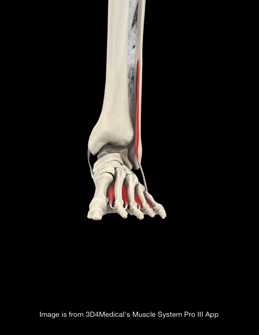
Overtightening of the ankle syndesmosis: is it really possible?
Tornetta P 3rd1, Spoo JE, Reynolds FA, Lee C.
http://www.ncbi.nlm.nih.gov/pubmed/11315776
J Bone Joint Surg Am. 2001 Apr;83-A(4):489-92.

Femur rotation
http://journals.lww.com/acsm-msse/Abstract/publishahead/Femur_Rotation_Increases_Patella_Cartilage_Stress.97824.aspx

Reader:
Hi there Dr Ivo and Dr Allen
I thought this article may interest you.
http://leonchaitow.com/2015/01/21/rediscovering-better-posture-a-foot-related-personal-saga/

http://www.mortonsfoot.com/pickingrightpci.html
This last paragraph/quote in particular caught my eye.
I was wondering what your opinion of this would be and wether you agree with it entirely?
Wenger et al (1989) suggest that, since flexible flat foot is generally a benign condition, it rarely requires treatment.

wreck method, squats ?
https://www.weckmethod.com/articles/improve-squatting-form-using-the-neutral-squat-technique

High performance high heels ?

oy. At $925 a pair, maybe she is the smart one (but we don’t think so). There is always an opportunist it seems.

“If I was to continue to [wear heels] in the same level as I did in my 20s, I would literally end up with super deformed, damaged feet,” said Singh, 36.
Traditionally, the structure of a high heel relies on a single metal shank running along the sole of the shoe providing a very stiff — and uncomfortable — platform that puts 75 percent of the pressure on the ball of the foot.
Singh and her team are trying to adjust the distribution of load so that the ball of the foot carries 50 percent of the weight and the heel carries the other 50 percent such that the impact — and with it, the pain — is lessened.

http://nypost.com/2015/05/03/these-high-performance-heels-wont-kill-your-feet/

One of our favorite (and biggest brained) people in the world !In his own words: “Gait is the most important biomarker for neurological integrity and is considered a quantifiable variable that might serve as a window of disease or functional n…

One of our favorite (and biggest brained) people in the world !
In his own words: “Gait is the most important biomarker for neurological integrity and is considered a quantifiable variable that might serve as a window of disease or functional neurological progression or attenuation.”

Prof. Carrick has had almost 4 decades of clinical experience in this gait knowledge area. His knowledge application base in this functional approach to human stance, balance and gait is world renown. He will share his clinical experience so that the learner will be able to gain a fluency in this exciting evidence based clinical area of expertise.
Check out this teaser 15" talk on his Amsterdam course coming up in June.

http://lp.carrickinstitute.org/advanced-human-gait/

Increase glute medius activity with weighted unilateral carries.

Looking for a way to increase functional-gait related gluteus medius activity, skill, endurance and strength ? Do not forget to implement the unilateral weighted “carries”.
The forward walking lunge and split squats are similar exercises that have differences in the eccentric phase, and both can be performed in the ipsilateral or contralateral carrying conditions. Contralateral walking lunges highly activate the gluteus medius. You can vary these challenges with unilateral overhead carry (the waiter’s carry) to help engage the shoulder, core and incorporate breathing skills.

http://journals.lww.com/…/DOES_THE_DUMBBELL_CARRYING_POSITI…

Running yourself into thyroid troubles ?

A few years ago we wrote about this:
“Are you running yourself into hormonal problems?”
here is the link: http://thegaitguys.tumblr.com/…/are-you-running-yourself-in…

Well, there has been some new research and one of our fav authors has brought things to present day research. Here is what Alex Hutchinson has to say and share with us all over as Sweat Science.

http://www.runnersworld.com/health/endurance-training-and-thyroid-function

Weighted Carries for glute training

Looking for a way to increase functional-gait related gluteus medius activity, skill, endurance and strength ? Do not forget to implement the unilateral weighted “carries”.
The forward walking lunge and split squats are similar exercises that have differences in the eccentric phase, and both can be performed in the ipsilateral or contralateral carrying conditions. Contralateral walking lunges highly activate the gluteus medius. You can vary these challenges with unilateral overhead carry (the waiter’s carry) to help engage the shoulder, core and incorporate breathing skills.

http://journals.lww.com/nsca-jscr/Abstract/publishahead/DOES_THE_DUMBBELL_CARRYING_POSITION_CHANGE_THE.96900.aspx

Mental training

“research findings that mental skills are underutilized by injured athletes in the 3 countries examined. More effort should be focused on educating and training athletes, coaches, and sports medicine professionals on the effectiveness of mental training in the injury rehabilitation context.”

http://journals.humankinetics.com/jsr-current-issue/jsr-volume-24-issue-2-may/athletes-use-of-mental-skills-during-sport-injury-rehabilitation

Shoes and Pronation and Injuries

Our brief (very) thoughts on the topic in Triathlete.com. We do not do much of this stuff anymore, our thoughts get so “cooked down” that they often lose context. But thankfully the main principle was conveyed pretty well here, albeit not in entirety.

We were asked about this topic many moons ago. We did not know this was published in January, until a patient brought it to our attention this week.

http://triathlon.competitor.com/2015/01/training/pronation-blame-injuries_111767

Beware of research data

Be aware of what you read.
We have had many people over the years criticize some of the articles we put up….“hey, the N on this study is 6 ! That isn’t a reliable study” etc.
As we have said many times, you can pick apart many studies. Few are comprehensive, many just look at a small piece, and as our link today eludes to, some are frauds and listed from “pay-for-play” publications. Some however are just so flawed that publication should never have occurred.
However, just because you have a problem with an article does necessarily mean to throw the baby out with the bath water. Good or bad, most valid articles have something good or bad to learn from. Sometimes they spark ideas in our minds, sometime they encourage thought, change or avoidance. This is the value of a valid journal article to us, the bigger picture, not because the study only looked at one aspect of a theory or hypothesis.

Here is an article that is raising Cane in the nutrition world and it sort of highlights some problems.

“Bohannon, a science journalist who also has a Ph.D., lays out how he carried out an elaborate hoax to expose just how easily bad nutrition science gets disseminated in the mainstream media. "You have to know how to read a scientific paper — and actually bother to do it,” he writes. For starters, as Bohannon explains in great detail, the study design itself was flawed — it had too few subjects, and the research measured too many factors, making it likelier that some random factor would appear to have statistical significance.

http://www.npr.org/sections/thesalt/2015/05/28/410313446/why-a-journalist-scammed-the-media-into-spreading-bad-chocolate-science

The cross over gait and achilles pathologies.

From our post earlier today:

Part 2: 

Considered another way, from the top down this time, if at the moment of heel contact the gmedius is delayed (as suggested in the study below from achilles pain), the pelvis is likely to drift laterally and this could cause a reactive inversion strategy of the rearfoot, and maybe even forefoot as well, as an instinctive measure to try and draw support beneath the laterally drifting body mass center of gravity. (This in essence sets up the “cross over gait” deployment strategy we have talked about here for years now).This too could cause a change in load to the achilles mechanism, resulting in tendonopathy thus putting one into a vicious cycle of achilles causing glute and then glute perpetuating altered strike and thus abnormal achilles loads . This is also a major cause of ankle inversion sprains, so be extra aware of this pattern. We see this all the time in practice, we hope you do as well. IF you haven’t already, If you wish to dive deeper, goto our blogwww.thegaitguys.tumblr.com and type in Cross Over Gait and it will get you going.
* Remember this, if you are spending time moving sideways, you are taking from time moving forwards, in the allotted amount of time given for the stance phase of gait. Yet, you will still move forwards, so one way around this is a premature heel rise (ie. speed up certain mechanical events) via premature plantarflexion mechanism loading (calf-achilles complex). Remember to also look for all the other reasons for premature heel rise (ie. loss of ankle rocker etc).

Franettovich Smith MM1, Honeywill C, Wyndow N, Crossley KM, Creaby MW. : Neuromotor control of gluteal muscles in runners with achilles tendinopathy.
Med Sci Sports Exerc. 2014 Mar;46(3):594-9.

______________

Part 1

The mighty Gluteus Medius, in all its glory!

Perhaps the delayed action of the gluteus medius allows an adductory moment of the pelvis, moving the center of gravity medially. This could conceivably place additional stress on the achilles tendon (via the lateral gastroc) to create more eversion of the foot from midstance onward.

“The results of the study demonstrate altered neuromuscular control of the GMED and GMED in runners with Achilles Tendonitis. During running, GMED typically activates before heel strike so as to stabilize the hip and the pelvis. In runners with Achilles Tendonitis, GMED is activated with a delay, which consequently might affect the kinematics of knee and ankle resulting in rear foot inversion. Similarly, GMAX is activated with a delay and for a shorter duration in runners with Achilles Tendonitis. GMAX is the primary hip extensor and via a kinetic chain, a decreased hip extension moment might be compensated by an increased ankle plantarflexion moment which could potentially increase the load on the Achilles tendon.”

Franettovich Smith MM1, Honeywill C, Wyndow N, Crossley KM, Creaby MW. : Neuromotor control of gluteal muscles in runners with achilles tendinopathy.
Med Sci Sports Exerc. 2014 Mar;46(3):594-9.

Post stroke spatiotemporal gait asymmetries.

This could be translated into your non-stroke patients gait. Making changes in their gait must be specific, accurate (what they need, not changes that you are seeing which you do not like), and consciously engrained through repetition. As with most things, awareness is the first step toward change. And making changes based on what you see is just asking them to create a new pattern on what was likely their compensation, not their problem. Never change what you see in someones gait without a complete physical examination first, what you see is how they are moving with their presenting problem(s).

“Asymmetry magnitudes need to exceed usual overground levels to reach conscious awareness. Therefore, it is proposed that the spatiotemporal asymmetry that is specific to each participant may need to be augmented beyond what he or she usually walks with in order to promote awareness of asymmetric gait patterns for long-term correction and learning.”
“Following stroke, spatiotemporal gait asymmetries persist into the chronic phases, despite the neuromuscular capacity to produce symmetric walking patterns. This persistence of gait asymmetry may be due to deficits in perception, as the newly established asymmetric gait pattern is perceived as normal.”

http://ptjournal.apta.org/…/2015/04/30/ptj.20140482.abstract

Walking meditation

A different perspective on walking.
When we give our gait retraining homework to clients, we ask that it is mindful and that each step is focused on the changes we have recommended. We ask that they notice the local changes and eventually the global manifestations of these gait alterations. This is key to making changes, and as this piece eludes to……there is something deeper cerebrally going on that can change other deeper aspects of your life.

How to Walk is the fourth title in Parallax’s popular Mindfulness Essentials Series of how-to titles by Zen Master Thich Nhat Hanh, introducing beginners and reminding seasoned practitioners of the essentials of mindfulness practice. Slow, concentrated walking while focusing on in- and out-breaths allows for a unique opportunity to be in the present. - See more at: 

http://www.parallax.org/how-to-walk-thich-nhat-hanh-mindfulness-essentials/

Isometrics for patellar tendonitis?We are familiar with different modes of exercise: isometric, isotonic and isokinetic. Isometric exercises have a physiological overflow of 10 degrees on each side of the point of application (ie; to do the exercise…

Isometrics for patellar tendonitis?

We are familiar with different modes of exercise: isometric, isotonic and isokinetic. Isometric exercises have a physiological overflow of 10 degrees on each side of the point of application (ie; to do the exercise at 20 degrees flexion, and you have strength gains from 10 to 30 degrees); isotonics and isokinetics, 15 degrees. Taking advantage of physiological overflow often allows us to bypass painful ranges of motion and still strengthen in that range of motion. 

In this study, they looked at immediate and 45 minute later pain reduction (not function) comparing isometric (max voluntary quadricep contraction) and isotonic (single leg decline squat) exercises. They also looked at cortical inhibition (via the cortico spinal tract) as a result of the exercises. 

Here is what they found: “A single resistance training bout of isometric contractions reduced tendon pain immediately for at least 45 min postintervention and increased MVIC. The reduction in pain was paralleled by a reduction in cortical inhibition, providing insight into potential mechanisms. Isometric contractions can be completed without pain for people with PT. The clinical implications are that isometric muscle contractions may be used to reduce pain in people with PT without a reduction in muscle strength.” These same results were not seen with the isotonic exercise. 

Did the decrease in pain result in the decrease in cortical inhibition (muscle contraction is inhibited across an inflamed joint: Rice, McNair 2010; Iles, Stokes 1987)? Was it a play on post isometric inhibition (most likely not, since this usually only lasts seconds to minutes post contraction) ? Or is there another mechanism at play here? There has been one other paper we found here, that shows cortical inhibition of quadriceps post isometric exercise. Time will tell. In the meantime, start using those multiple angle isometrics!

The Gait Guys

Rio E, Kidgell D, Purdam C, Gaida J, Moseley GL, Pearce AJ, Cook J.Isometric exercise induces analgesia and reduces inhibition in patellar tendinopathy Br J Sports Med. 2015 May 15. pii: bjsports-2014-094386. doi: 10.1136/bjsports-2014-094386. [Epub ahead of print]

http://www.anatomy-physiotherapy.com/28-systems/musculoskeletal/lower-extremity/knee/1163-isometric-exercises-in-patellar-tendinopathy

The mighty Gluteus Medius, in all its glory!Perhaps the delayed action of the gluteus medius allows an adductory moment of the pelvis, moving the center of gravity medially. This could conceivably place additional stress on the achilles tendon  (via…

The mighty Gluteus Medius, in all its glory!

Perhaps the delayed action of the gluteus medius allows an adductory moment of the pelvis, moving the center of gravity medially. This could conceivably place additional stress on the achilles tendon  (via the lateral gastroc) to create more eversion of the foot from midstance on

“The results of the study demonstrate altered neuromuscular control of the GMED and GMED in runners with Achilles Tendonitis. During running, GMED typically activates before heel strike so as to stabilize the hip and the pelvis. In runners with Achilles Tendonitis, GMED is activated with a delay, which consequently might affect the kinematics of knee and ankle resulting in rear foot inversion. Similarly, GMAX is activated with a delay and for a shorter duration in runners with Achilles Tendonitis. GMAX is the primary hip extensor and via a kinetic chain, a decreased hip extension moment might be compensated by an increased ankle plantarflexion moment which could potentially increase the load on the Achilles tendon.”

Franettovich Smith MM1, Honeywill C, Wyndow N, Crossley KM, Creaby MW. : Neuromotor control of gluteal muscles in runners with achilles tendinopathy.
Med Sci Sports Exerc. 2014 Mar;46(3):594-9.

tumblr_np3dt87iAK1qhko2so1_540.png
tumblr_np3dt87iAK1qhko2so2_540.png

The Mighty Interossei

By request of one of our readers (Thank you Richard S), we were asked to “dig up” some information about the interossei. After scouring the literature, we turned up an interesting paper, talking about their anatomy. 

Of interesting note, the paper found extensive connections of the musculature with the surrounding fascia (talk about myofascial meridians!) as well as a fairly consistent slip of the peroneus longus which contributes to the 1st dorsal interossei. This is important considering the peroneus fires from midstance on, as do the interossei (and other foot intrinsics). Perhaps (since as the fore foot is extending in late midstance and pre swing) it assists in descending the head of the 1st metatarsal and resisting extension (contracting eccentrically) of the metatarsal phalangeal joints, helping to maintain stability of the fore foot for push off. 

“The extensive connections among the interossei indicate that they could be important stabilisers of the foot during those times when rigidity is required. The pull of the interossei is transformed across the tarsometatarsal joints by means of their attachment to the ligamentous meshwork. Thus they will act upon the tarsometatarsal joints. Crossing those joints on their plantar aspect, the interossei are well placed to assist in resisting extension. Even though their close attachment proximal to the joints creates a short lever arm and therefore relative inefficiency as flexors when weight is borne on the ball of the foot (MacConaill, 1949), the large mass of the combined interossei probably indicates that they do have a significant role in resisting extension at these joints. Also, the shapes of the tarsometatarsal joint surfaces restrict angular motion.”

Definitely a good read and available FREE full text online here

PAUL J. KALINt AND BRUCE ELLIOT HIRSCH: The origins and function of the interosseous muscles of the foot  J. Anat. (1987), 152, pp. 83-91 

Abdominal Activity and GaitWe came across this cool study today, after a well educated patient asked about abdominal activity during gait.Here is the bottom line:low level activity in the rectus abdominis and external oblique throughout the gait cyc…

Abdominal Activity and Gait

We came across this cool study today, after a well educated patient asked about abdominal activity during gait.

Here is the bottom line:
low level activity in the rectus abdominis and external oblique throughout the gait cycle, more concentrated activity of the internal oblique at initial contact/loading response (heel strike).

This makes sense, since the external oblique occupies more real estate and has a larger cross sectional area; it most likely has a role in stabilization both in rotational like emoticon planes as well as the saggital plane (Z). Perhaps the action of the internal oblique at initial contact is to assist in external rotation of the pelvis on the stance phase leg, as the the opposite leg goes into swing?

“Cluster analysis identified two patterns of activity for the internal oblique, external oblique and rectus abdominis muscles. In the lumbar erector spinae, three patterns of activity were observed. In most instances, the patterns observed for each muscle differed in the magnitude of the activation levels. In rectus abdominis and external oblique muscles, the majority of subjects had low levels of activity (<5.0% of a maximum voluntary contraction) that were relatively constant throughout the stride cycle. In the internal oblique and the erector spinae muscles, more distinct bursts of activity were observed, most often close to foot-strike. The different algorithms used for the cluster analysis yielded similar results and a discriminant function analysis provided further evidence to support the patterns observed”

Clin Biomech (Bristol, Avon). 2002 Mar;17(3):177-84.
Abdominal and erector spinae muscle activity during gait: the use of cluster analysis to identify patterns of activity.
White SG1, McNair PJ.

Forefoot Varus Anyone?Forefoot varus appears to move the center of gravity medially while walking. Nothing earthshaking here, but nice to see the support of the literature.&ldquo;The most medial CoP of the row and CoP% detected increased medial CoP …

Forefoot Varus Anyone?

Forefoot varus appears to move the center of gravity medially while walking. Nothing earthshaking here, but nice to see the support of the literature.

“The most medial CoP of the row and CoP% detected increased medial CoP deviation in FV ≥ 8°, and may be applied to other clinical conditions where rearfoot angle and CoP of the array after initial heel contact cannot detect significant differences.”

We will be talking about foot types this week on onlinece.com; Wednesday 8 EST, 7 CST, 6MST, 5 PST Biomechanics 314. Hope to see you there!

J Formos Med Assoc. 2015 May 5. pii: S0929-6646(15)00132-1. doi: 10.1016/j.jfma.2015.03.004. [Epub ahead of print]
Analysis of medial deviation of center of pressure after initial heel contact in forefoot varus.

picture from: http://forums.teamestrogen.com/showthread.php?t=46901

tumblr_nozmpqndk41qhko2so1_540.png
tumblr_nozmpqndk41qhko2so2_500.png

Clinical tidbit:

Heel pain in kids and adolescents? Have you considered Sever’s disease?

Apophysitis of the calcaneal apophysis is the most common cause of heel pain in adloscents and accounts for 8% of all pediatric overuse injuries! An apophysitis occurs (an injury involving a “pulling away” of bone from the tendons attachment site) because the strength of the tendon exceeds the strength of attachment of the tendon to the bone. It is most common in activites llike running, jumping and plantar flexion.

Gillespie H. Osteochondroses and apophyseal injuries of the foot in the young athlete. Curr Sports Med Rep 2010;9(5):265-268.

Wilson JC, Rodenburg RE. Apophysitis of the lower extremities. Contemp Pediatr 2011;28(6):38-46.

tumblr_nozmmaOxAI1qhko2so1_500.png
tumblr_nozmmaOxAI1qhko2so2_540.png

Ahh yes, the lumbricals. 

One of our favorite muscles. And here it is in a recent paper! This one is for all you fellow foot geeks : )

Perhaps the FDL (which fires slightly earlier than the FHL) and FHL (which fires slightly later and longer) at loading response, slowing pronation and setting the stage for lumbrical function from midstance to terminal stance/preswing (flexion at the metatarsal phalangeal joint (it would have to be eccentric, if you think about this from a closed chain perspective) and extension (actually compression) of the proximal interphalangeal joints.

“The first lumbrical arose as two muscle bellies from both the tendon of the FDL and the tendinous slip of the FHL in 83.3 %, and as one muscle belly from the tendon of the FDL or the tendinous slip of the FHL in 16.7 %. These two muscle bellies subsequently merged to form the muscle belly of the first lumbrical. The second lumbrical arose from the tendinous slips of the FHL for the second and third toes as well as the tendon of the FDL in all specimens. The third lumbrical arose from the tendinous slips of the FHL for the third and fourth toes in 69.7 %, and the fourth lumbrical arose from the tendinous slip of the FHL for the fourth toe in 18.2 %. Some deep muscle fibers of the fourth lumbrical arose from the tendinous slip of the FHL for the second toe in 4.5 %, for the third toe in 28.8 %, and for the fourth toe in 15.2 %.”

Hur MS1, Kim JH, Gil YC, Kim HJ, Lee KS. New insights into the origin of the lumbrical muscles of the foot: tendinous slip of the flexor hallucis longus muscle. Surg Radiol Anat. 2015 May 12. [Epub ahead of print]